Copyright
©The Author(s) 2021.
World J Gastroenterol. May 7, 2021; 27(17): 1973-1992
Published online May 7, 2021. doi: 10.3748/wjg.v27.i17.1973
Published online May 7, 2021. doi: 10.3748/wjg.v27.i17.1973
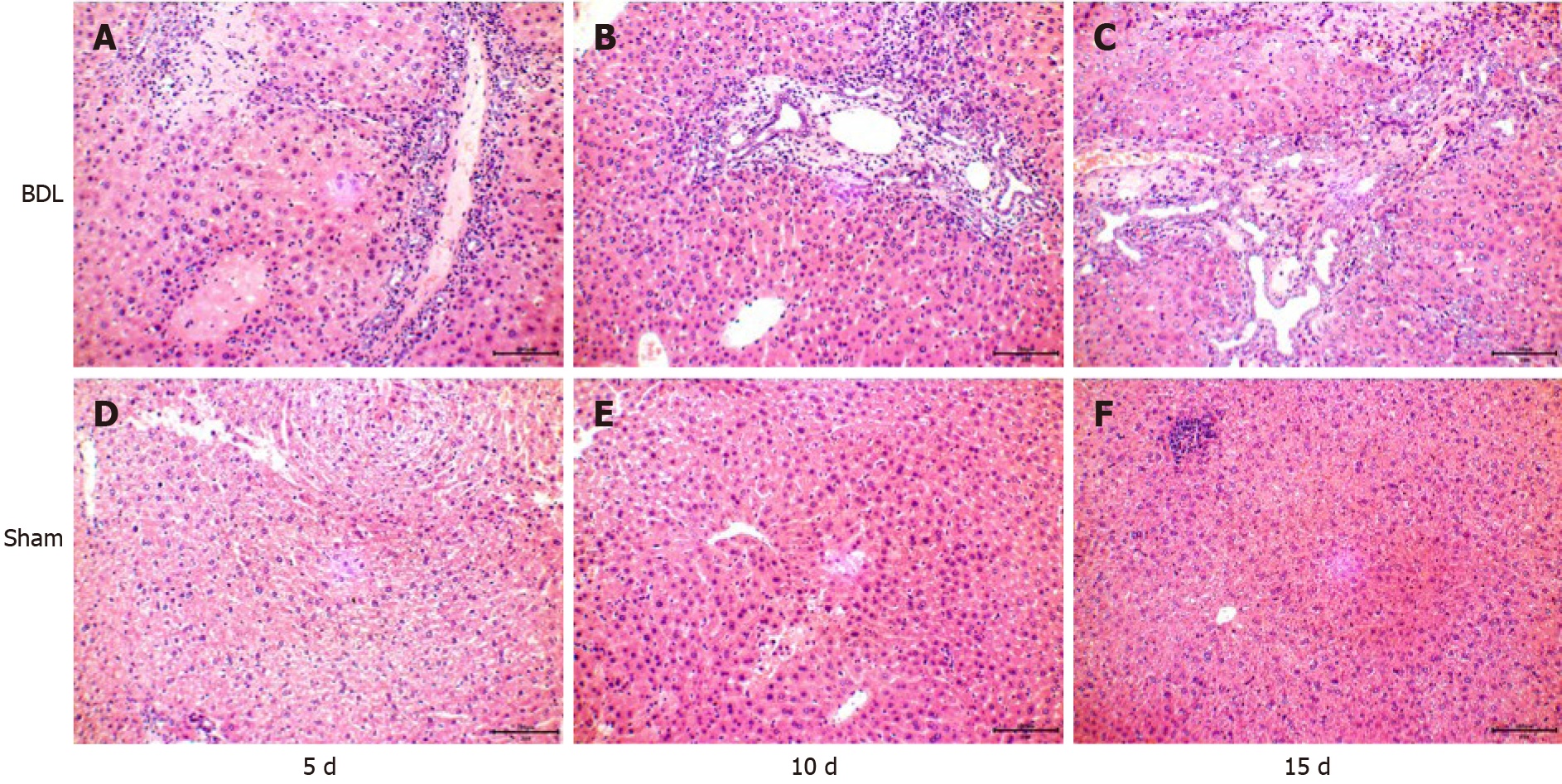
Figure 3 Liver tissues of mice in the bile duct ligation group and sham group were observed under an optical microscope (hematoxylin-eosin stain, × 400).
A-C: 5, 10 and 15 d after surgery in the bile duct ligation group, respectively; D-F: 5, 10 and 15 d after surgery in the sham group, respectively. BDL: Bile duct ligation.
- Citation: Dong XH, Dai D, Yang ZD, Yu XO, Li H, Kang H. S100 calcium binding protein A6 and associated long noncoding ribonucleic acids as biomarkers in the diagnosis and staging of primary biliary cholangitis. World J Gastroenterol 2021; 27(17): 1973-1992
- URL: https://www.wjgnet.com/1007-9327/full/v27/i17/1973.htm
- DOI: https://dx.doi.org/10.3748/wjg.v27.i17.1973


