Copyright
©The Author(s) 2021.
World J Gastroenterol. Apr 21, 2021; 27(15): 1655-1663
Published online Apr 21, 2021. doi: 10.3748/wjg.v27.i15.1655
Published online Apr 21, 2021. doi: 10.3748/wjg.v27.i15.1655
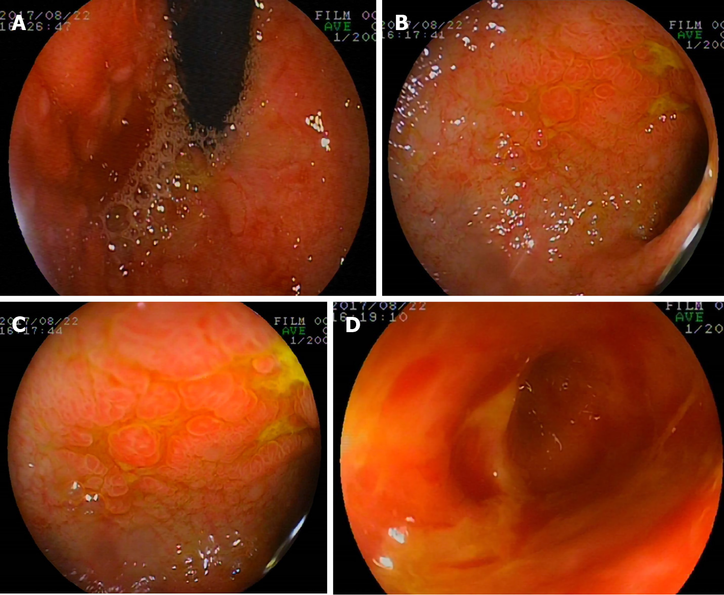
Figure 2 Gastroscopic findings.
A gastroscope was inserted into the horizontal part of the duodenum. Hyperemia, edema, lymphoid hyperplasia, focal ulceration, and pseudotumor formation were seen in the stomach and duodenum. A: Gastric fundus; B and C: Duodenal bulb; D: Dilatation of proximal duodenum.
- Citation: Yang QH, Ma XP, Dai DL, Bai DM, Zou Y, Liu SX, Song JM. Gastrointestinal cytomegalovirus disease secondary to measles in an immunocompetent infant: A case report. World J Gastroenterol 2021; 27(15): 1655-1663
- URL: https://www.wjgnet.com/1007-9327/full/v27/i15/1655.htm
- DOI: https://dx.doi.org/10.3748/wjg.v27.i15.1655


