Copyright
©The Author(s) 2021.
World J Gastroenterol. Apr 14, 2021; 27(14): 1419-1434
Published online Apr 14, 2021. doi: 10.3748/wjg.v27.i14.1419
Published online Apr 14, 2021. doi: 10.3748/wjg.v27.i14.1419
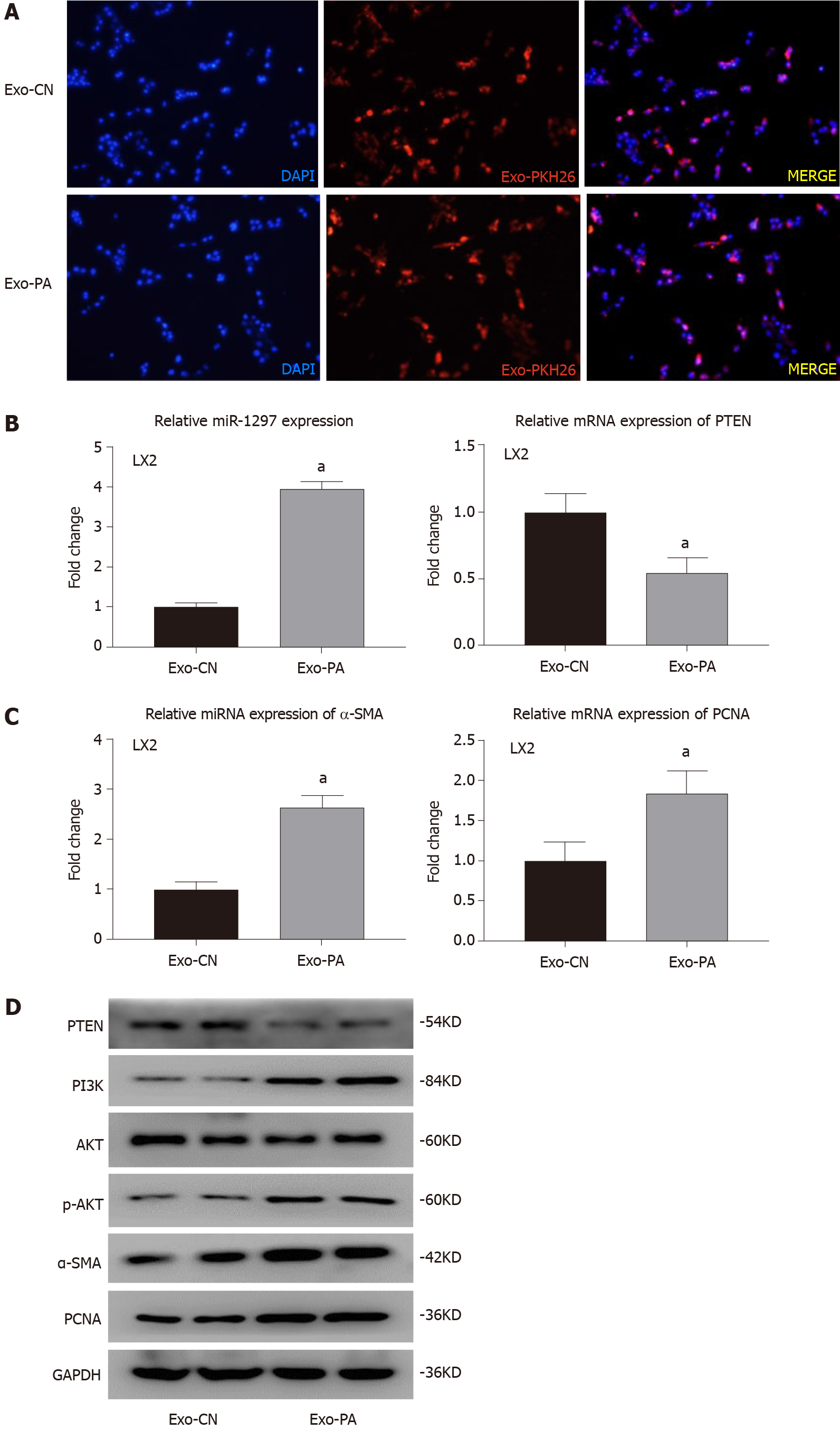
Figure 6 Lipotoxic hepatocyte-secreted exosomes could transfer exosomal microRNA-1297 to hepatic stellate cells and contribute to hepatic stellate cell activation through the PTEN/PI3K/AKT pathway.
A: The PKH-26 stained exosomes were absorbed by LX2 cells observed by a fluorescence microscope; B: The relative microRNA expression of miR-1297 and mRNA expression of PTEN were assessed by quantitative real-time PCR after exosomes derived from vehicle control or palmitic acid (Exo-CN or Exo-PA) treatment for 48 h. miR-1297 mimics of negative controls (mi-NC): 1.00 ± 0.12 vs miR1297 mimics (mi-miR): 3.95 ± 0.18, PTEN mi-NC: 1.00 ± 0.13 vs mi-miR: 0.54 ± 0.12; C: The relative mRNA expression of α-SMA and PCNA were assessed by quantitative real-time PCR after Exo-CN or Exo-PA treatment for 48 h. α-SMA mi-NC 1.00 ± 0.14 vs mi-miR 2.63 ± 0.23, PCNA mi-NC 1.00 ± 0.23 vs mi-miR 1.85 ± 0.23; D: Protein levels of α-SMA, PCNA, PTEN, PI3K, AKT and p-AKT in LX2 cells were assessed by western blot after Exo-CN or Exo-PA treatment for 48 h; E: Immunofluorescence staining was performed to evaluate the activation of LX2 cells after Exo-CN or Exo-PA treatment for 48 h; mi-NC: 1.00 ± 0.26 vs mi-miR: 1.68 ± 0.21; F: Ethynyl-20-deoxyuridine (EdU) staining was performed to evaluate the proliferation of LX2 cells after Exo-CN or Exo-PA treatment for 48 h, mi-NC: 0.21 ± 0.04 vs mi-miR: 0.41 ± 0.07. Statistical significance: aP < 0.05. DAPI: 4´6-Diamidino-2-phenylindole; Exo-CN: Exosomes from control vehicle; Exo-PA: Exosomes from palmitic acid.
- Citation: Luo X, Luo SZ, Xu ZX, Zhou C, Li ZH, Zhou XY, Xu MY. Lipotoxic hepatocyte-derived exosomal miR-1297 promotes hepatic stellate cell activation through the PTEN signaling pathway in metabolic-associated fatty liver disease. World J Gastroenterol 2021; 27(14): 1419-1434
- URL: https://www.wjgnet.com/1007-9327/full/v27/i14/1419.htm
- DOI: https://dx.doi.org/10.3748/wjg.v27.i14.1419


