Copyright
©The Author(s) 2021.
World J Gastroenterol. Mar 21, 2021; 27(11): 990-1005
Published online Mar 21, 2021. doi: 10.3748/wjg.v27.i11.990
Published online Mar 21, 2021. doi: 10.3748/wjg.v27.i11.990
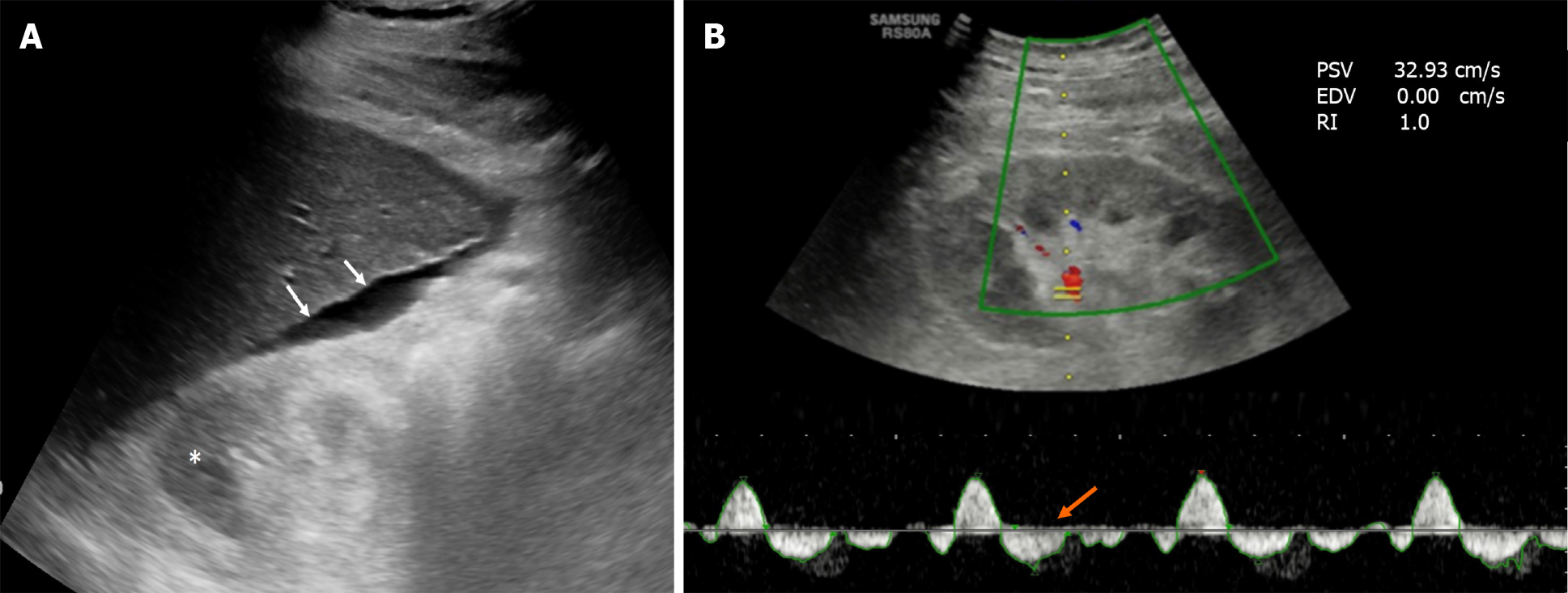
Figure 2 Ultrasonographic image of a 65-year-old diabetic patient with liver cirrhosis and chronic kidney disease.
A: The liver outline is irregular (white arrows) and there is ascites around it. The right kidney is small and the parenchymal echogenicity is increased with loss of corticomedullary differentiation (asterisk), suggesting chronic kidney disease; B: Doppler sonogram of the same kidney showed reversal of diastolic flow (orange arrow) with absent end-diastolic velocity, indicating very high resistance vessels.
- Citation: Kumar R, Priyadarshi RN, Anand U. Chronic renal dysfunction in cirrhosis: A new frontier in hepatology. World J Gastroenterol 2021; 27(11): 990-1005
- URL: https://www.wjgnet.com/1007-9327/full/v27/i11/990.htm
- DOI: https://dx.doi.org/10.3748/wjg.v27.i11.990


