Copyright
©The Author(s) 2020.
World J Gastroenterol. Dec 7, 2020; 26(45): 7263-7271
Published online Dec 7, 2020. doi: 10.3748/wjg.v26.i45.7263
Published online Dec 7, 2020. doi: 10.3748/wjg.v26.i45.7263
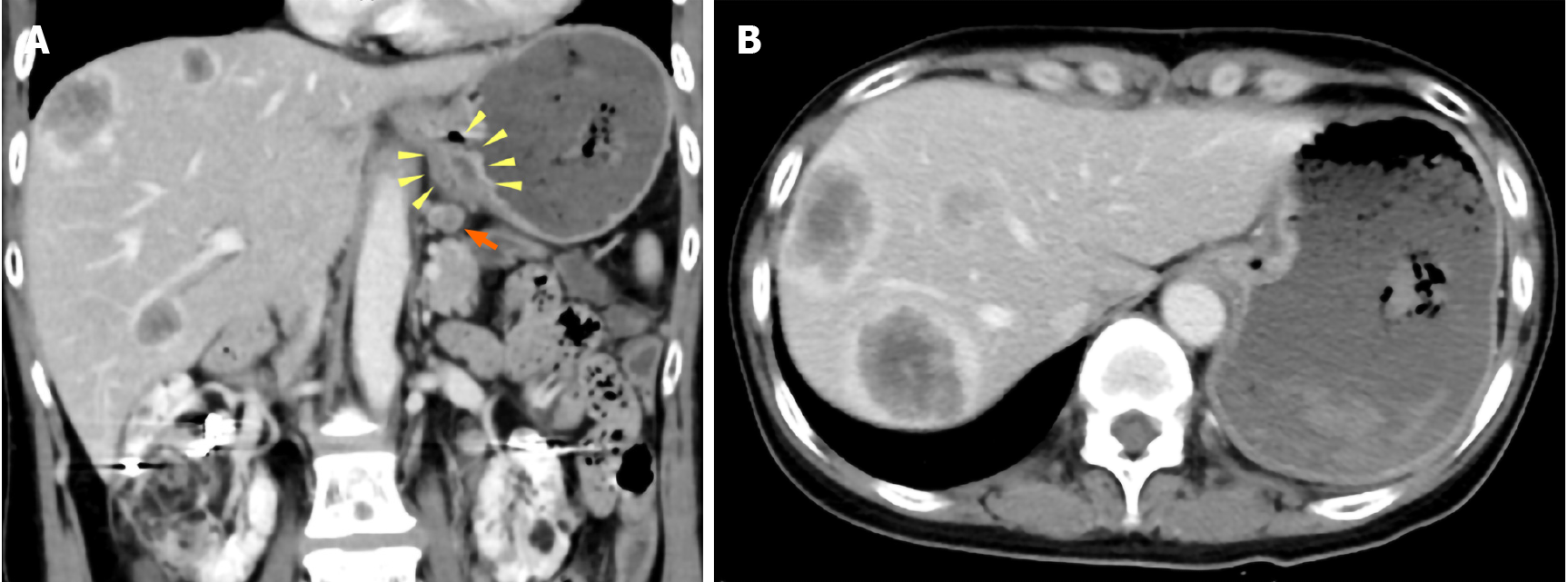
Figure 1 Abdominal contrast-enhanced computed tomography images.
A: Wall thickness from the esophagogastric junction to the cardia (yellow arrowhead) and enlarged lymph nodes near the lesser curvature of the stomach (orange arrow); B: Multiple ring-enhanced tumors are observed in the liver.
- Citation: Ishida N, Miyazu T, Tamura S, Suzuki S, Tani S, Yamade M, Iwaizumi M, Osawa S, Hamaya Y, Shinmura K, Sugimura H, Miura K, Furuta T, Sugimoto K. Tuberous sclerosis patient with neuroendocrine carcinoma of the esophagogastric junction: A case report. World J Gastroenterol 2020; 26(45): 7263-7271
- URL: https://www.wjgnet.com/1007-9327/full/v26/i45/7263.htm
- DOI: https://dx.doi.org/10.3748/wjg.v26.i45.7263


