Copyright
©The Author(s) 2020.
World J Gastroenterol. Nov 14, 2020; 26(42): 6689-6697
Published online Nov 14, 2020. doi: 10.3748/wjg.v26.i42.6689
Published online Nov 14, 2020. doi: 10.3748/wjg.v26.i42.6689
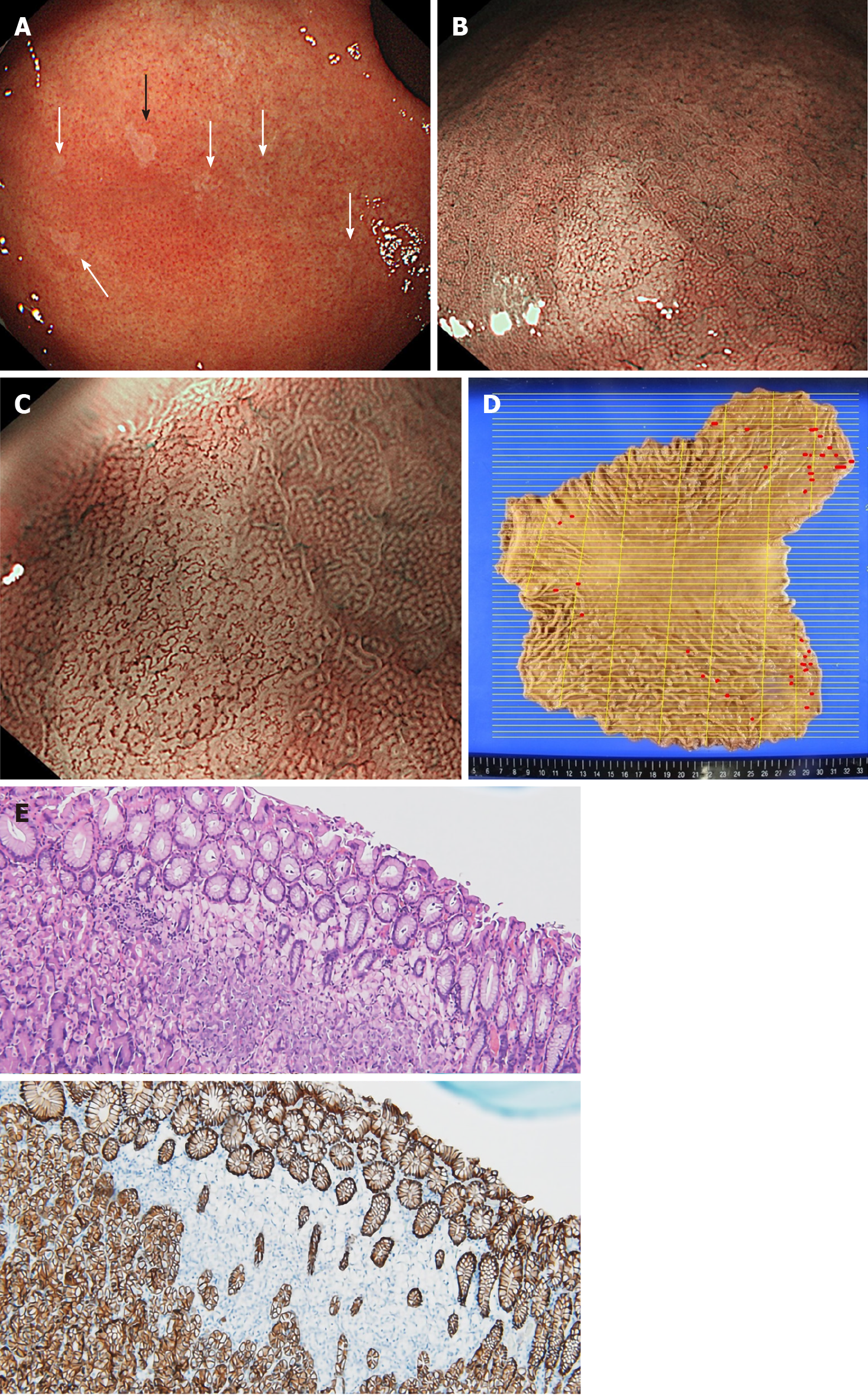
Figure 4 Representative images obtained from esophagogastroduodenoscopy and pathological findings in Case 3.
A: Multiple small pale lesions were observed mainly at the greater curvature of the gastric body in esophagogastroduodenoscopy (white and black arrows); B: Clearly isolated whitish areas were detected by non-magnifying narrow band imaging (NBI). The image is the lesion indicated by the black arrow in (A); C: Magnifying NBI detected wavy microvessels inside the lesions; D: A gastrectomy mapping study revealed 36 signet ring cell carcinoma (SRCC) foci in the entire gastric mucosa. Red lines indicate SRCC foci; E: Hematoxylin and eosin staining (upper panel) and immunohistochemistry for E-cadherin (lower panel) of the lesion. Loss of immunoreactivity at SRCC foci was confirmed.
- Citation: Hirakawa M, Takada K, Sato M, Fujita C, Hayasaka N, Nobuoka T, Sugita S, Ishikawa A, Mizukami M, Ohnuma H, Murase K, Miyanishi K, Kobune M, Takemasa I, Hasegawa T, Sakurai A, Kato J. Case series of three patients with hereditary diffuse gastric cancer in a single family: Three case reports and review of literature. World J Gastroenterol 2020; 26(42): 6689-6697
- URL: https://www.wjgnet.com/1007-9327/full/v26/i42/6689.htm
- DOI: https://dx.doi.org/10.3748/wjg.v26.i42.6689


