Copyright
©The Author(s) 2020.
World J Gastroenterol. Nov 14, 2020; 26(42): 6556-6571
Published online Nov 14, 2020. doi: 10.3748/wjg.v26.i42.6556
Published online Nov 14, 2020. doi: 10.3748/wjg.v26.i42.6556
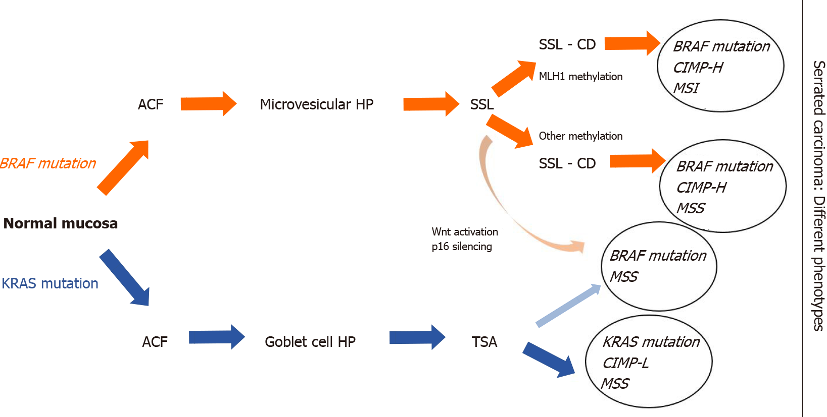
Figure 2 Outline of the schematic serrated pathway progression.
In red color we indicate the steps of transformation of BRAF-mutated serrated lesions. BRAF mutations and hypermethylation lead to transformation of aberrant crypt foci to microvesicular hyperplastic polyp then to sessile serrated lesions (SSLs). Methylation and loss of key tumor suppressor genes such as p16 and MLH1 are the key points in SSLs’ progression to serrated adenocarcinoma. In blue color we indicate KRAS mutations in traditional serrated adenomas (TSAs), which showed MGMT hypermethylation, but not MLH1 promoter hypermethylation. In light red shading we indicate a non-MLH1 mutating SSL, which could progress to a TSA and ultimately develop into a BRAF-mutated microsatellite stability tumor. ACF: Aberrant crypt foci; HP: Hyperplastic polyp; SSL: Sessile serrated lesion; SSL-CD: Sessile serrated lesion with cytological dysplasia; TSA: Traditional serrated adenoma; CIMP: CpG island hypermethylator phenotype; CIMP-H: CIMP-high; CIMP-L: CIMP-low; MSI: Microsatellite instability; MSS: Microsatellite stability.
- Citation: Peruhova M, Peshevska-Sekulovska M, Krastev B, Panayotova G, Georgieva V, Konakchieva R, Nikolaev G, Velikova TV. What could microRNA expression tell us more about colorectal serrated pathway carcinogenesis? World J Gastroenterol 2020; 26(42): 6556-6571
- URL: https://www.wjgnet.com/1007-9327/full/v26/i42/6556.htm
- DOI: https://dx.doi.org/10.3748/wjg.v26.i42.6556


