Copyright
©The Author(s) 2020.
World J Gastroenterol. Nov 7, 2020; 26(41): 6361-6377
Published online Nov 7, 2020. doi: 10.3748/wjg.v26.i41.6361
Published online Nov 7, 2020. doi: 10.3748/wjg.v26.i41.6361
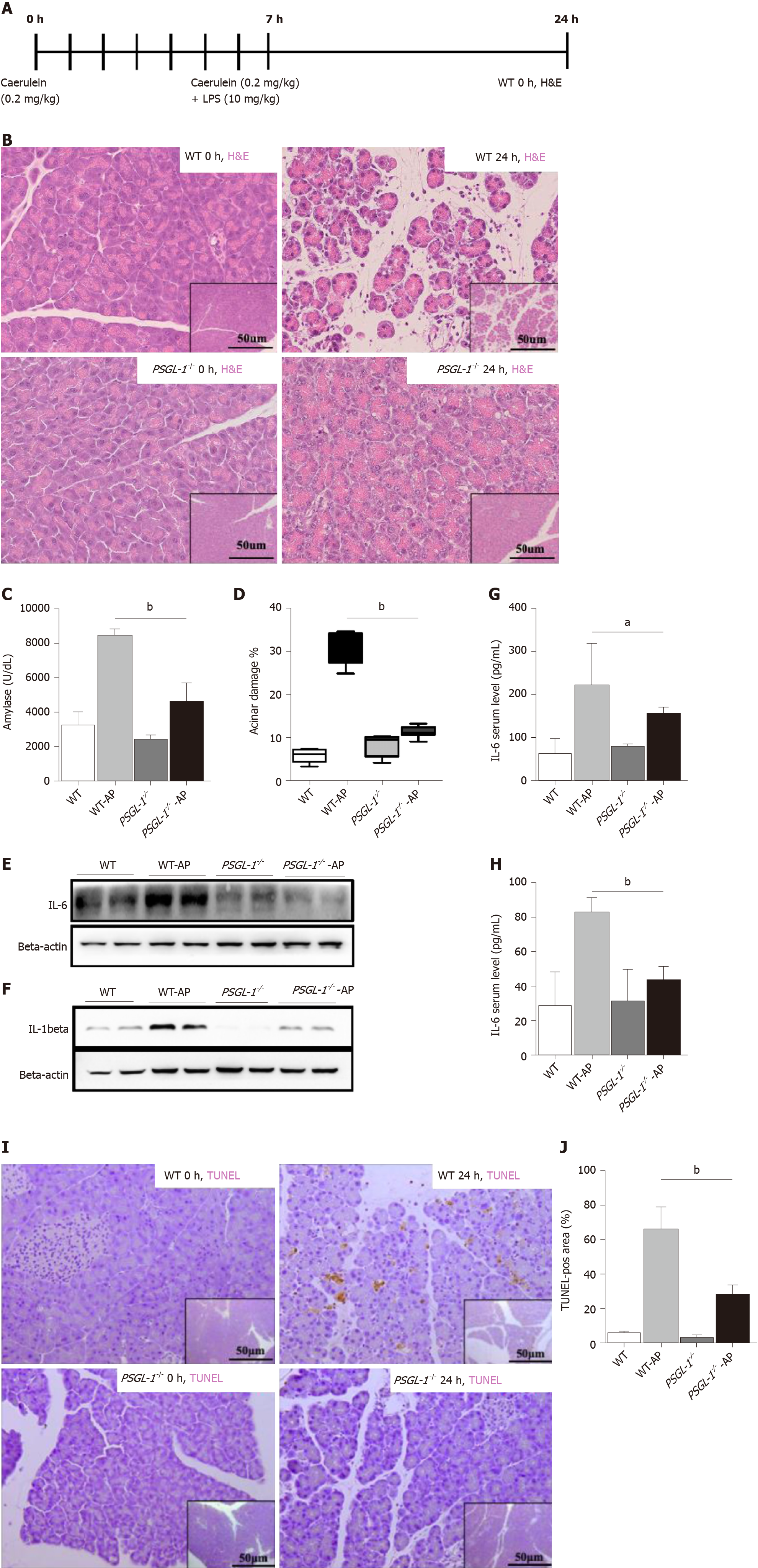
Figure 2 P-selectin glycoprotein ligand 1 deficiency alleviates caerulein-mediated inflammatory response and acinar damage.
A: Schematic diagram of the induction of acute pancreatitis (AP) showing the frequency of caerulein and lipopolysaccharide injections as well as the sampling time points; B: Hematoxylin-eosin staining of the pancreas of wild-type (upper panels) and P-selectin glycoprotein ligand 1 (PSGL-1)-/- (lower panels) mice 0 h and 24 h after treatment with caerulein; C: Boxplots showing the expression of amylase (n = 4); D: Boxplots showing quantification of acinar cell damage (n = 4); E: Expression of IL-6 in the pancreas of wild-type and PSGL-1-/- mice; F: Expression of IL-1beta in the pancreas of wild-type and PSGL-1-/- mice; G: Expression of IL-6 in sera of wild-type and PSGL-1-/- mice (n = 4); H: Expression of IL-1beta in sera of wild-type and PSGL-1-/- mice(n = 4); I: Transferase-mediated dUTP-biotin nick end labeling (TUNEL) assay for apoptosis in the pancreas; J: Boxplots showing quantification of the TUNEL assays (n = 4). aP < 0.05; bP < 0.001, Student's t-test. H&E: Hematoxylin-eosin; TUNEL: Transferase-mediated dUTP-biotin nick end labeling; PSGL-1: P-selectin glycoprotein ligand 1; AP: Acute pancreatitis; WT: Wild type.
- Citation: Zhang X, Zhu M, Jiang XL, Liu X, Liu X, Liu P, Wu XX, Yang ZW, Qin T. P-selectin glycoprotein ligand 1 deficiency prevents development of acute pancreatitis by attenuating leukocyte infiltration. World J Gastroenterol 2020; 26(41): 6361-6377
- URL: https://www.wjgnet.com/1007-9327/full/v26/i41/6361.htm
- DOI: https://dx.doi.org/10.3748/wjg.v26.i41.6361


