Copyright
©The Author(s) 2020.
World J Gastroenterol. Oct 28, 2020; 26(40): 6241-6249
Published online Oct 28, 2020. doi: 10.3748/wjg.v26.i40.6241
Published online Oct 28, 2020. doi: 10.3748/wjg.v26.i40.6241
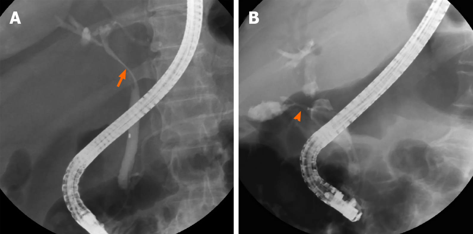
Figure 1 Cholangiography of patients with Mirizzi syndrome.
A: Patient without a cholecystocholedochal fistula. Eccentric compression (orange arrow) of the common bile duct is observed and a short part of the cystic duct is opacified; B: Patient with a cholecystocholedochal fistula (orange arrowhead). A contrast opacified gallbladder without the typical spiral and corkscrew-like cystic duct opacification is shown.
- Citation: Wu CH, Liu NJ, Yeh CN, Wang SY, Jan YY. Predicting cholecystocholedochal fistulas in patients with Mirizzi syndrome undergoing endoscopic retrograde cholangiopancreatography. World J Gastroenterol 2020; 26(40): 6241-6249
- URL: https://www.wjgnet.com/1007-9327/full/v26/i40/6241.htm
- DOI: https://dx.doi.org/10.3748/wjg.v26.i40.6241


