Copyright
©The Author(s) 2020.
World J Gastroenterol. Sep 7, 2020; 26(33): 5008-5021
Published online Sep 7, 2020. doi: 10.3748/wjg.v26.i33.5008
Published online Sep 7, 2020. doi: 10.3748/wjg.v26.i33.5008
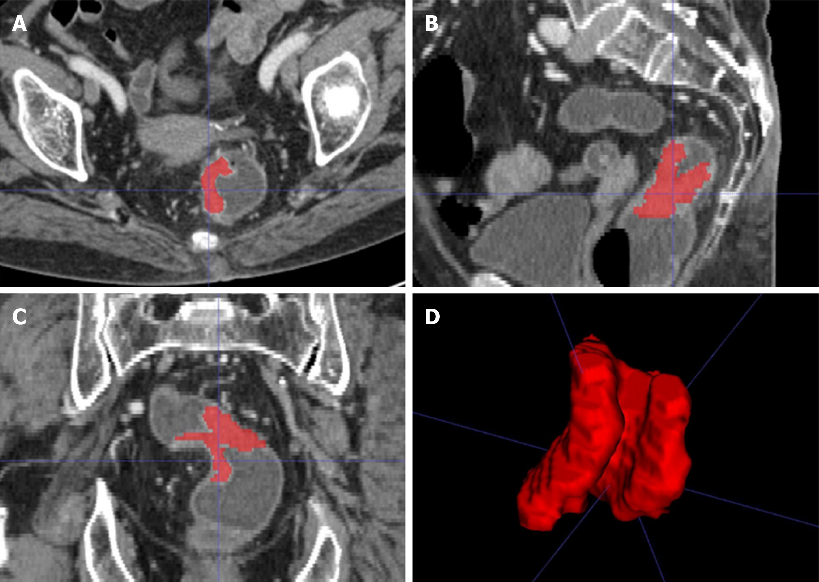
Figure 2 A 58-year-old woman with rectal cancer.
A-C: Representative manual segmentation of the whole lesion in the axial, sagittal, and coronal enhanced computed tomography images. Red lines represent the delineations of the regions of interest used to derive the radiomic features; D: Three-dimensional volumetric reconstruction of the segmented lesion.
- Citation: Li M, Zhu YZ, Zhang YC, Yue YF, Yu HP, Song B. Radiomics of rectal cancer for predicting distant metastasis and overall survival. World J Gastroenterol 2020; 26(33): 5008-5021
- URL: https://www.wjgnet.com/1007-9327/full/v26/i33/5008.htm
- DOI: https://dx.doi.org/10.3748/wjg.v26.i33.5008


