Copyright
©The Author(s) 2020.
World J Gastroenterol. Jan 21, 2020; 26(3): 366-374
Published online Jan 21, 2020. doi: 10.3748/wjg.v26.i3.366
Published online Jan 21, 2020. doi: 10.3748/wjg.v26.i3.366
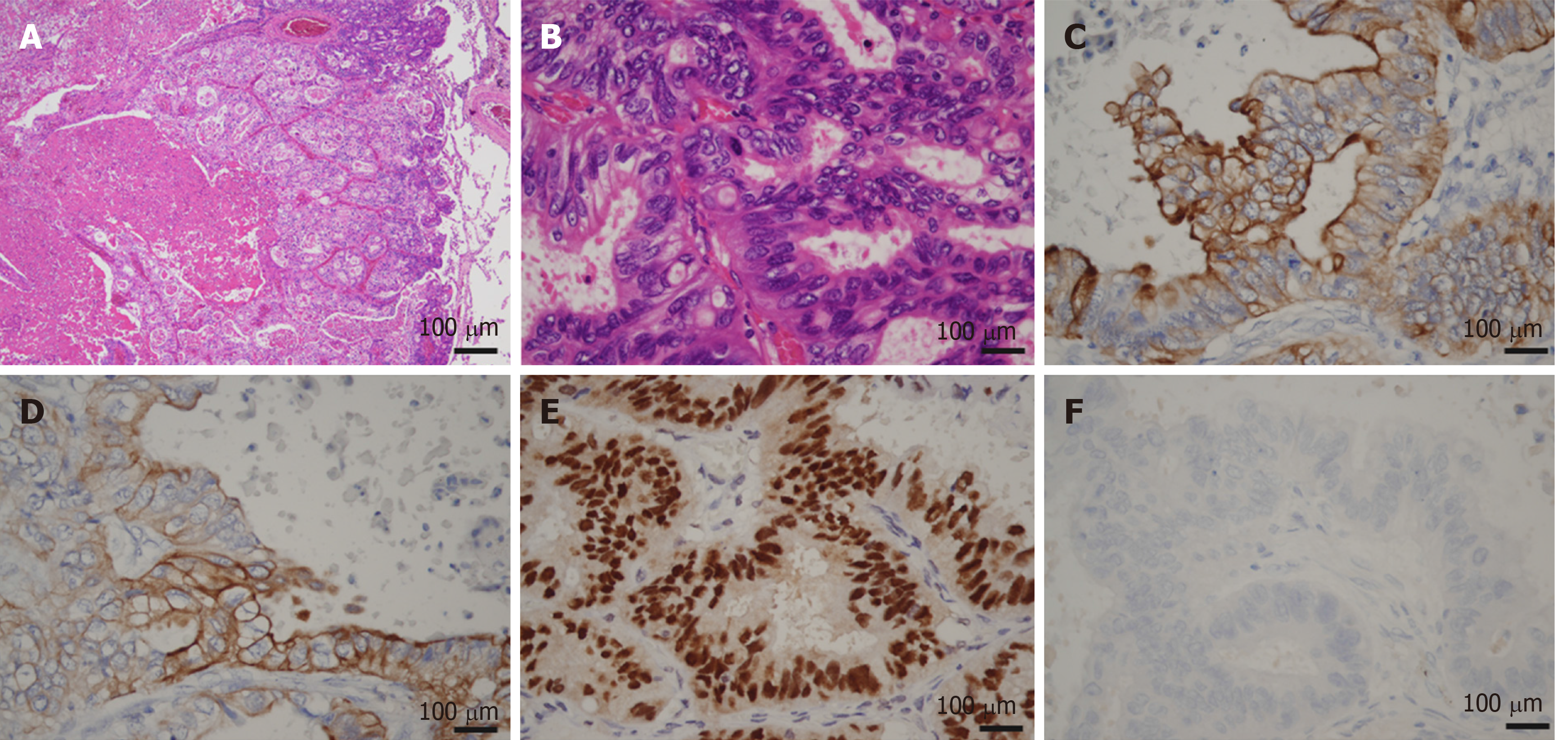
Figure 8 Microscopic images of lung metastasis.
A and B: Histologic appearance showed a well-demarcated nodule with marked necrosis in the center of the tumor (A: magnification × 20, B: magnification × 400). At high power magnification, there were eosinophilic, tall, columnar cells with nuclear pseudostratification. C-E: Immunohistological staining showed that the tumor cells were positive for CK7 (Panel C), CK20 (Panel D) and CDX2 (Panel E); F: Immunohistological staining showed that the tumor cells were negative for TTF-1.
- Citation: Nam NH, Taura K, Kanai M, Fukuyama K, Uza N, Maeda H, Yutaka Y, Chen-Yoshikawa TF, Muto M, Uemoto S. Unexpected metastasis of intraductal papillary neoplasm of the bile duct without an invasive component to the brain and lungs: A case report. World J Gastroenterol 2020; 26(3): 366-374
- URL: https://www.wjgnet.com/1007-9327/full/v26/i3/366.htm
- DOI: https://dx.doi.org/10.3748/wjg.v26.i3.366


