Copyright
©The Author(s) 2020.
World J Gastroenterol. Jul 21, 2020; 26(27): 3938-3951
Published online Jul 21, 2020. doi: 10.3748/wjg.v26.i27.3938
Published online Jul 21, 2020. doi: 10.3748/wjg.v26.i27.3938
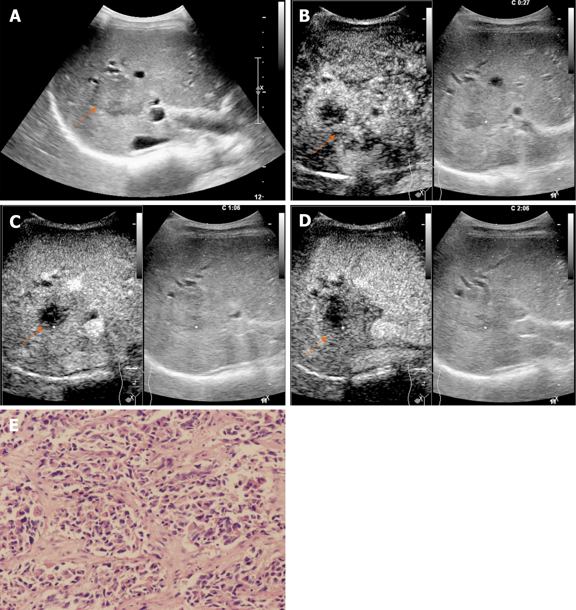
Figure 2 A 54-year-old female patient with a lesion categorized as LR-M.
A: Conventional grayscale ultrasound detected a hypoechoic nodule (arrow) 3.6 cm in diameter in the right lobe of the liver; B: Rim arterial phase hyperenhancement (APEH) (arrow) in the arterial phase was demonstrated by contrast-enhanced ultrasound; C and D: No washout (arrow) was observed in the early portal phase (by 60 s), and no marked washout (arrow) was observed by 126 s after SonoVue injection. This lesion was designated as LR-M because of rim APEH in the arterial phase; E: Poorly differentiated intrahepatic cholangiocarcinoma was confirmed by histopathology (hematoxylin and eosin staining, × 200).
- Citation: Huang JY, Li JW, Ling WW, Li T, Luo Y, Liu JB, Lu Q. Can contrast enhanced ultrasound differentiate intrahepatic cholangiocarcinoma from hepatocellular carcinoma? World J Gastroenterol 2020; 26(27): 3938-3951
- URL: https://www.wjgnet.com/1007-9327/full/v26/i27/3938.htm
- DOI: https://dx.doi.org/10.3748/wjg.v26.i27.3938


