Copyright
©The Author(s) 2020.
World J Gastroenterol. Jul 21, 2020; 26(27): 3929-3937
Published online Jul 21, 2020. doi: 10.3748/wjg.v26.i27.3929
Published online Jul 21, 2020. doi: 10.3748/wjg.v26.i27.3929
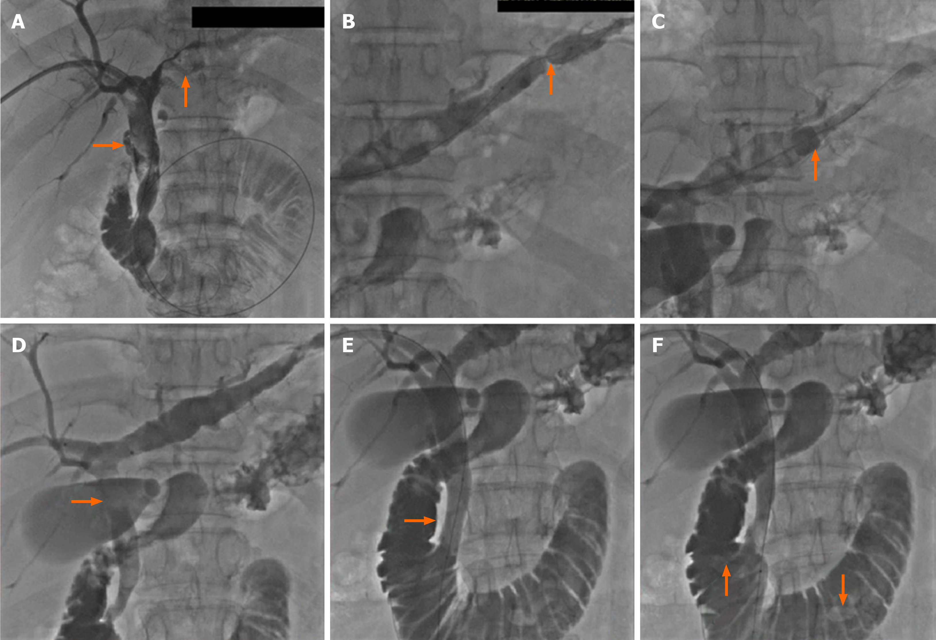
Figure 2 Cholangiography.
A: Cholangiography revealed the location, size, and number of the intrahepatic and common bile duct stones; B: The left bile duct with stones was dilated with a balloon catheter in presence of stenosis; C: The intrahepatic bile duct stone was pushed with a dilated Fogarty catheter; D: Stone was pushed into common bile duct; E: The papilla was dilated; F: Stones in the common bile duct were pushed into the duodenum.
- Citation: Liu B, Cao PK, Wang YZ, Wang WJ, Tian SL, Hertzanu Y, Li YL. Modified percutaneous transhepatic papillary balloon dilation for patients with refractory hepatolithiasis. World J Gastroenterol 2020; 26(27): 3929-3937
- URL: https://www.wjgnet.com/1007-9327/full/v26/i27/3929.htm
- DOI: https://dx.doi.org/10.3748/wjg.v26.i27.3929


