Copyright
©The Author(s) 2020.
World J Gastroenterol. Jun 28, 2020; 26(24): 3484-3494
Published online Jun 28, 2020. doi: 10.3748/wjg.v26.i24.3484
Published online Jun 28, 2020. doi: 10.3748/wjg.v26.i24.3484
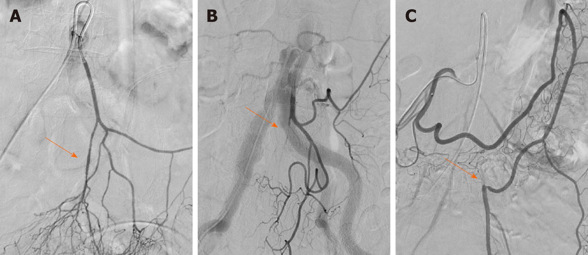
Figure 2 Demonstration of lesions in the inferior mesenteric artery and branches of the inferior mesenteric artery on digital subtraction angiography.
The arrows represent the lesion sites including stenosis and occlusion. A: Superior rectal artery stenosis with plaque; B: Superior rectal artery occlusion; C: Inferior mesenteric artery trunk occlusion. The distal blood supply came from left colic artery compensation for the superior mesenteric artery.
- Citation: Zhang C, Li A, Luo T, Li Y, Li F, Li J. Evaluation of characteristics of left-sided colorectal perfusion in elderly patients by angiography. World J Gastroenterol 2020; 26(24): 3484-3494
- URL: https://www.wjgnet.com/1007-9327/full/v26/i24/3484.htm
- DOI: https://dx.doi.org/10.3748/wjg.v26.i24.3484


