Copyright
©The Author(s) 2020.
World J Gastroenterol. Jun 14, 2020; 26(22): 2967-2986
Published online Jun 14, 2020. doi: 10.3748/wjg.v26.i22.2967
Published online Jun 14, 2020. doi: 10.3748/wjg.v26.i22.2967
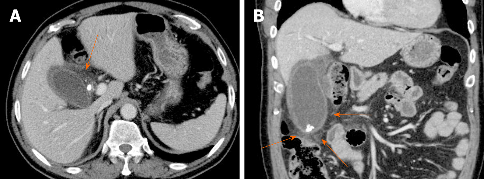
Figure 12 Acute cholecystitis.
A, B: Axial (A) and coronal (B) computed tomography scans of gallbladder showing tensile distension, diffuse mural thickening/edema, and pericholecystic fat stranding (arrows); impacted stone of cystic duct and gallstones present.
- Citation: Yu MH, Kim YJ, Park HS, Jung SI. Benign gallbladder diseases: Imaging techniques and tips for differentiating with malignant gallbladder diseases. World J Gastroenterol 2020; 26(22): 2967-2986
- URL: https://www.wjgnet.com/1007-9327/full/v26/i22/2967.htm
- DOI: https://dx.doi.org/10.3748/wjg.v26.i22.2967


