
















©The Author(s) 2020.
World J Gastroenterol. May 7, 2020; 26(17): 2012-2029
Published online May 7, 2020. doi: 10.3748/wjg.v26.i17.2012
Published online May 7, 2020. doi: 10.3748/wjg.v26.i17.2012
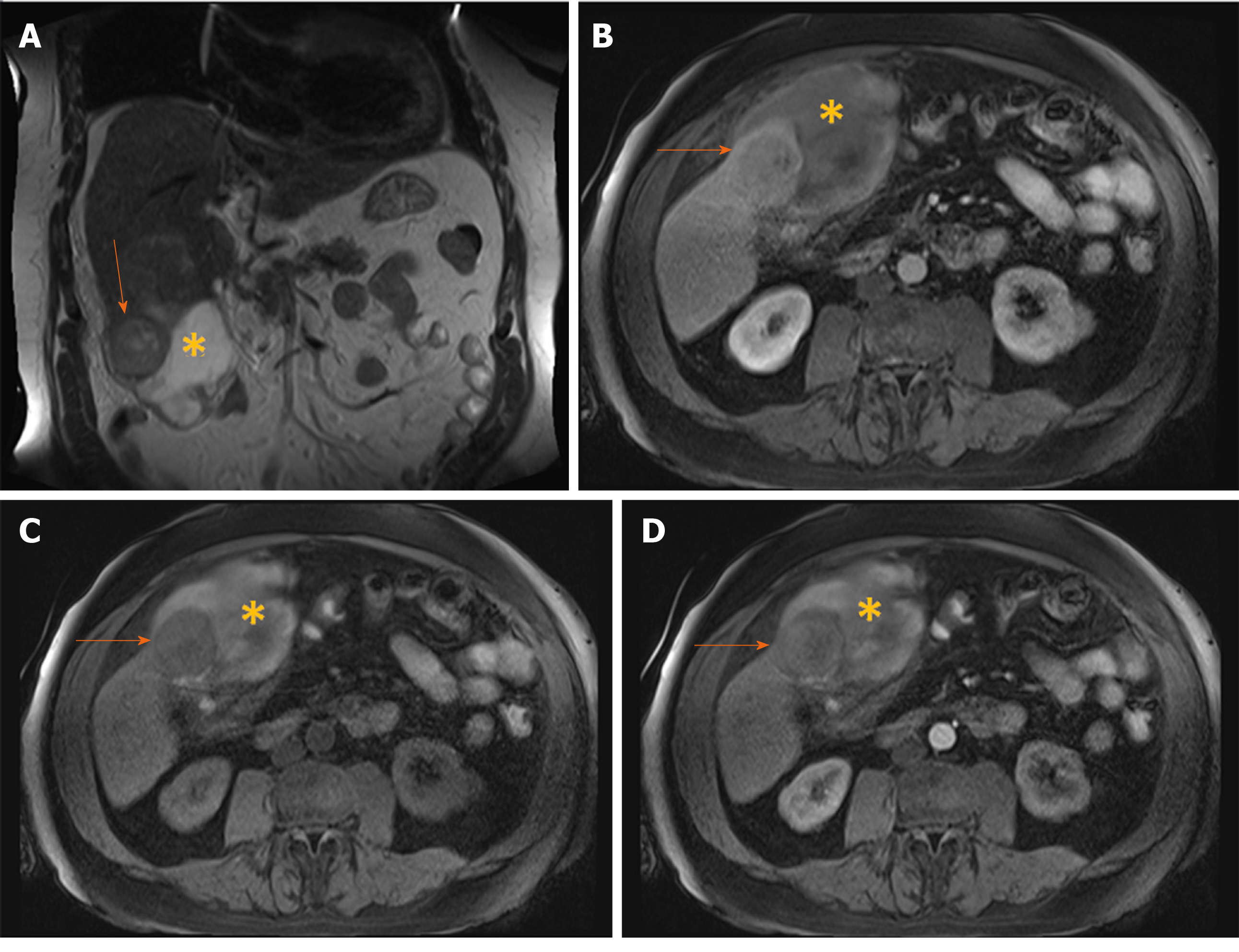
Figure 14 Hemorrhagic hepatocellular carcinoma in 70-year old man with cirrhosis presenting with acute abdominal pain.
A: Coronal T2-weighted image shows a nodular tumor (arrows) in the subcapsular location in segment V, and subhepatic hematoma (asterix); B: Axial T1-weighted FS image shows hypointense tumor (arrows) and hyperintense content of the hematoma (asterix); C and D: Arterial phase displays only subtle hypervascularity in the part of the tumor (arrows) depicted on this section (C) with washout in portal venous phase (D), and hematoma (asterix) located anteriorly.
- Citation: Kovac JD, Milovanovic T, Dugalic V, Dumic I. Pearls and pitfalls in magnetic resonance imaging of hepatocellular carcinoma. World J Gastroenterol 2020; 26(17): 2012-2029
- URL: https://www.wjgnet.com/1007-9327/full/v26/i17/2012.htm
- DOI: https://dx.doi.org/10.3748/wjg.v26.i17.2012