
















©The Author(s) 2020.
World J Gastroenterol. May 7, 2020; 26(17): 2012-2029
Published online May 7, 2020. doi: 10.3748/wjg.v26.i17.2012
Published online May 7, 2020. doi: 10.3748/wjg.v26.i17.2012
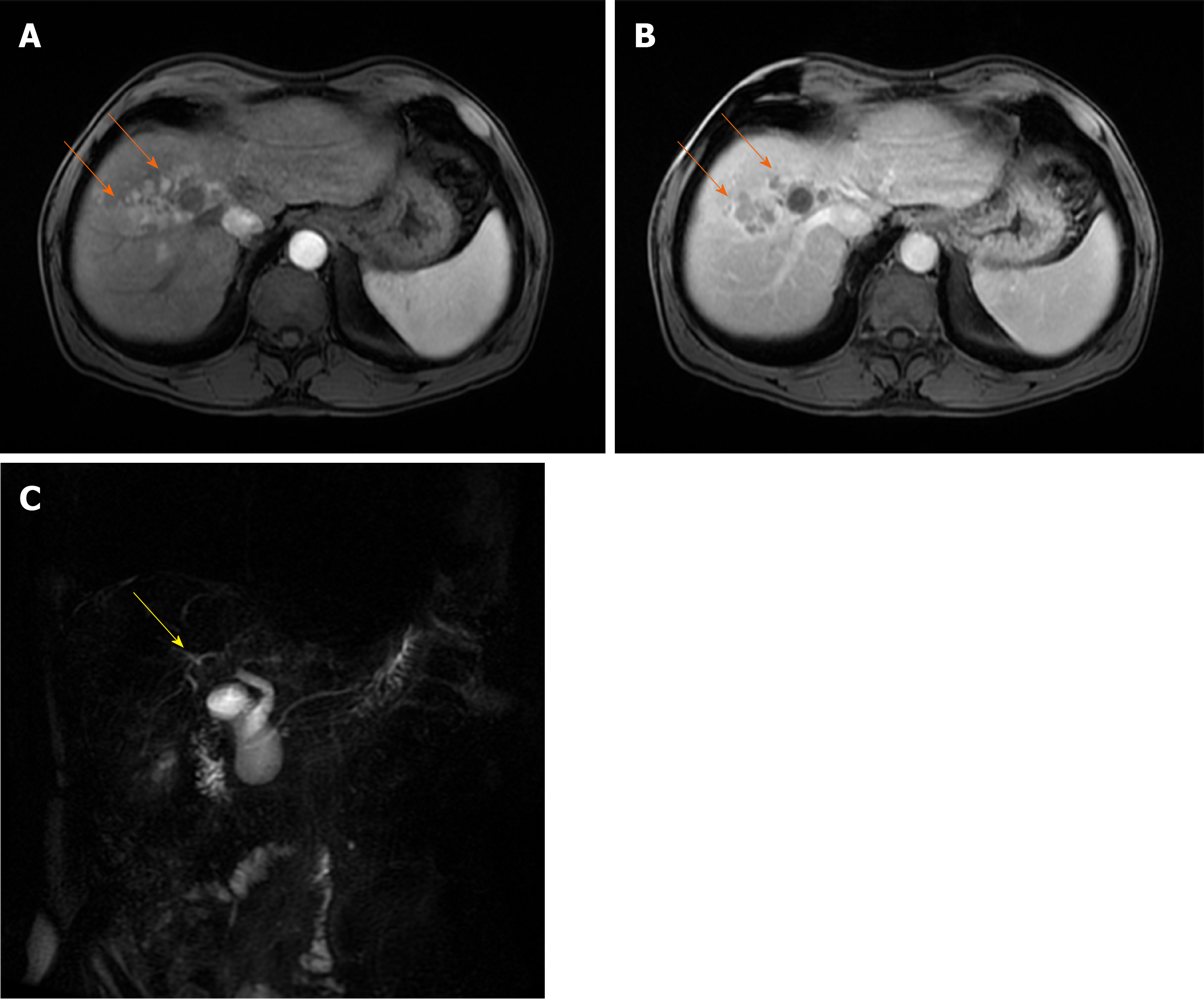
Figure 12 Infiltrative hepatocellular carcinoma in 63-year old man with intra-bile tumor growth.
A and B: On arterial phase image ill-defined hypervascular lesion (arrows) in segment VIII is seen (A) with washout in portal venous phase (B); C: Intra-bile tumor growth is better depicted on coronal magnetic resonance cholangiopancreatography image seen as defect in the lumen right hepatic duct and its posterior branch (yellow arrow).
- Citation: Kovac JD, Milovanovic T, Dugalic V, Dumic I. Pearls and pitfalls in magnetic resonance imaging of hepatocellular carcinoma. World J Gastroenterol 2020; 26(17): 2012-2029
- URL: https://www.wjgnet.com/1007-9327/full/v26/i17/2012.htm
- DOI: https://dx.doi.org/10.3748/wjg.v26.i17.2012