Copyright
©The Author(s) 2020.
World J Gastroenterol. Apr 14, 2020; 26(14): 1564-1579
Published online Apr 14, 2020. doi: 10.3748/wjg.v26.i14.1564
Published online Apr 14, 2020. doi: 10.3748/wjg.v26.i14.1564
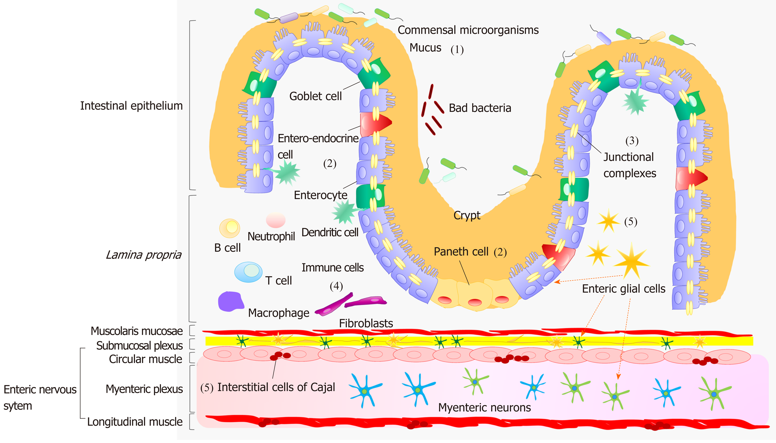
Figure 1 Diagram showing the morphology of intestinal epithelial barrier and neuromuscular compartment.
(1) The intestinal mucosa is covered by a hydrated gel, consisting mainly of mucins secreted by goblet cells The outer mucus layer provides a habitat for commensal microorganisms, while the inner mucus layer acts as a physical barrier preventing the penetration of microorganisms and other noxious agents into bowel tissues; (2) The epithelium includes: enterocytes that act as a selective physical barrier and regulatenutrient absorption, goblet cells, entero-endocrine cells that release intestinal hormones or peptides, and Paneth cells that regulate microbial populations and protect neighboring stem cells; (3) Junctional complexes confer mechanical strength to the intestinal epithelial barrier and regulate paracellular permeability; (4) The lamina propria, besides containing a number of innate and adaptive immune cells that respond to the insults with the secretion of inflammatory mediators, such as prostaglandins, histamine, and cytokines, is characterized by an intricate network of fibroblasts playing a key role in the proliferation of intestinal epithelium; and (5) Enteric glial cells, a cellular component of the enteric nervous system, are associated with both submucosal and myenteric neurons and are located also in proximity of epithelial cells. They coordinate signal propagation from and to myenteric neurons and epithelial cells, thus regulating bowel motility as well as the secretory and absorptive functions of enteric epithelium; interstitial cells of Cajal are the source of the electrical slow waves responsible for the transmission of excitation to the neighboring smooth muscle cells.
- Citation: D’Antongiovanni V, Pellegrini C, Fornai M, Colucci R, Blandizzi C, Antonioli L, Bernardini N. Intestinal epithelial barrier and neuromuscular compartment in health and disease. World J Gastroenterol 2020; 26(14): 1564-1579
- URL: https://www.wjgnet.com/1007-9327/full/v26/i14/1564.htm
- DOI: https://dx.doi.org/10.3748/wjg.v26.i14.1564


