Copyright
©The Author(s) 2020.
World J Gastroenterol. Mar 28, 2020; 26(12): 1340-1351
Published online Mar 28, 2020. doi: 10.3748/wjg.v26.i12.1340
Published online Mar 28, 2020. doi: 10.3748/wjg.v26.i12.1340
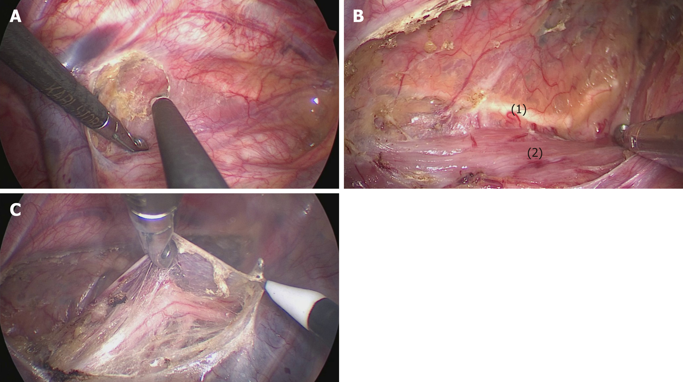
Figure 2 The mediastinal pleura was dissected on both sides.
A: The posterior mediastinal pleura above the arch of the azygos vein was dissected along the spine; B: The thoracic duct was fully exposed and carefully preserved, protecting the esophageal arteries on the dorsal side; C: The right vagus nerve was exposed by cutting the pleura toward the right subclavian artery. (1) Thoracic duct; (2) Esophagus.
- Citation: Chen WS, Zhu LH, Li WJ, Tu PJ, Huang JY, You PL, Pan XJ. Novel technique for lymphadenectomy along left recurrent laryngeal nerve during thoracoscopic esophagectomy. World J Gastroenterol 2020; 26(12): 1340-1351
- URL: https://www.wjgnet.com/1007-9327/full/v26/i12/1340.htm
- DOI: https://dx.doi.org/10.3748/wjg.v26.i12.1340


