Copyright
©The Author(s) 2019.
World J Gastroenterol. Feb 21, 2019; 25(7): 744-776
Published online Feb 21, 2019. doi: 10.3748/wjg.v25.i7.744
Published online Feb 21, 2019. doi: 10.3748/wjg.v25.i7.744
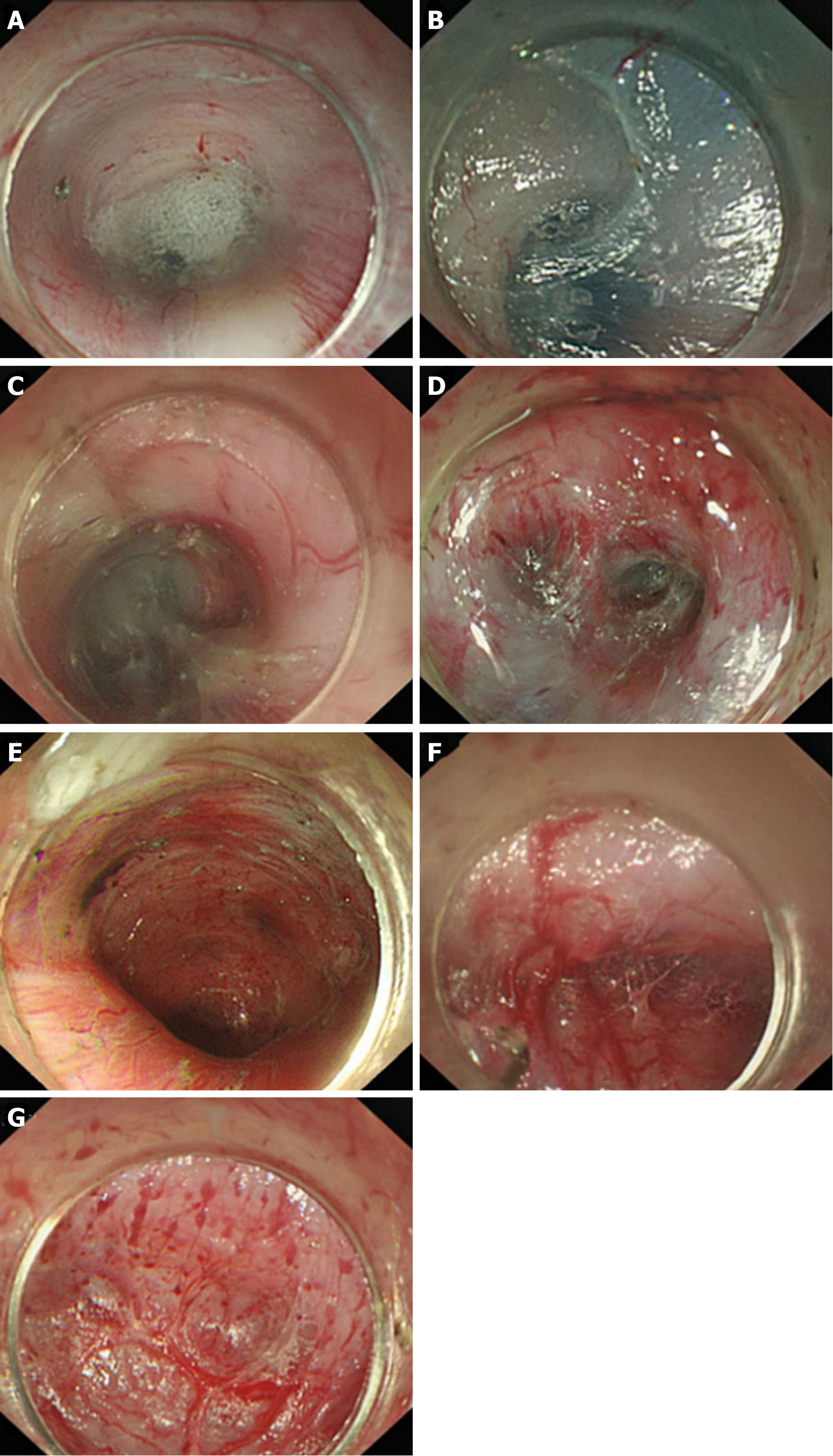
Figure 9 Anatomical landmark in the tunnel from the lower oesophagus to the cardia.
A: Grid-like blood vessels in the cardia; B: Crescent-like structure visible at the proximal cardia; C: Ampulla-like structure appearing after entering the crescent-like structure; D: Branching vessels with bulky vascular roots in the ampulla-like structure; E: Tunnel below the cardia, showing a steep downward form; F: Stubby and multi-branched vessels below the cardia; G: Beadlike vessels below the cardia.
- Citation: Chai NL, Li HK, Linghu EQ, Li ZS, Zhang ST, Bao Y, Chen WG, Chiu PW, Dang T, Gong W, Han ST, Hao JY, He SX, Hu B, Hu B, Huang XJ, Huang YH, Jin ZD, Khashab MA, Lau J, Li P, Li R, Liu DL, Liu HF, Liu J, Liu XG, Liu ZG, Ma YC, Peng GY, Rong L, Sha WH, Sharma P, Sheng JQ, Shi SS, Seo DW, Sun SY, Wang GQ, Wang W, Wu Q, Xu H, Xu MD, Yang AM, Yao F, Yu HG, Zhou PH, Zhang B, Zhang XF, Zhai YQ. Consensus on the digestive endoscopic tunnel technique. World J Gastroenterol 2019; 25(7): 744-776
- URL: https://www.wjgnet.com/1007-9327/full/v25/i7/744.htm
- DOI: https://dx.doi.org/10.3748/wjg.v25.i7.744


