Copyright
©The Author(s) 2019.
World J Gastroenterol. Feb 7, 2019; 25(5): 622-631
Published online Feb 7, 2019. doi: 10.3748/wjg.v25.i5.622
Published online Feb 7, 2019. doi: 10.3748/wjg.v25.i5.622
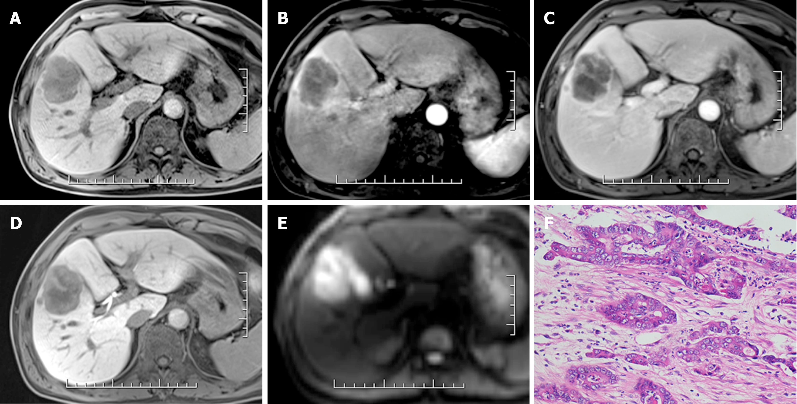
Figure 3 Pathologically confirmed intrahepatic cholangiocarcinoma in a 47-year-old man.
A: A 53.2-mm hypointense mass is seen on the precontrast image; B: The image shows rim-like hyperenhancement in the arterial phase; C: Peripheral “washout” in the portal venous phase; D: Targetoid appearance in the hepatobiliary phase; and E: Target sign in diffusion-weighted imaging; F: The mass was confirmed as intrahepatic cholangiocarcinoma at 200 × magnification after hematoxylin-eosin staining.
- Citation: Zhang T, Huang ZX, Wei Y, Jiang HY, Chen J, Liu XJ, Cao LK, Duan T, He XP, Xia CC, Song B. Hepatocellular carcinoma: Can LI-RADS v2017 with gadoxetic-acid enhancement magnetic resonance and diffusion-weighted imaging improve diagnostic accuracy? World J Gastroenterol 2019; 25(5): 622-631
- URL: https://www.wjgnet.com/1007-9327/full/v25/i5/622.htm
- DOI: https://dx.doi.org/10.3748/wjg.v25.i5.622


