Copyright
©The Author(s) 2019.
World J Gastroenterol. Feb 7, 2019; 25(5): 567-583
Published online Feb 7, 2019. doi: 10.3748/wjg.v25.i5.567
Published online Feb 7, 2019. doi: 10.3748/wjg.v25.i5.567
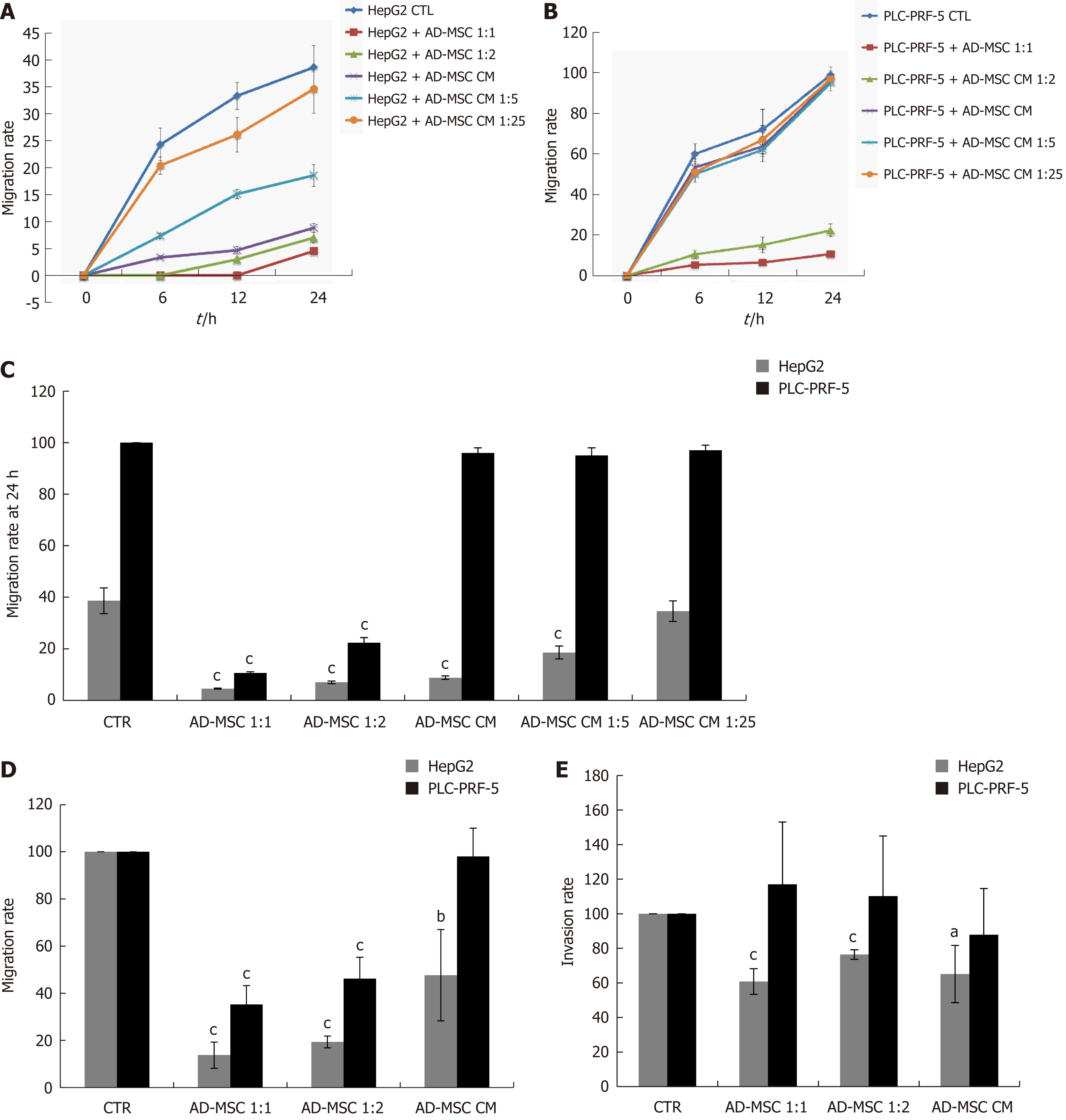
Figure 4 Effect of adipose derived mesenchymal stem cells and their conditioned media on of hepatocellular carcinoma cell migration and invasion.
HCC cells (2 × 10⁵) were seeded in six- well co-culture plates in the presence or absence of ADMSCs (at ADSMCs: HCC ratio of 1:1 or 1:2) or ADMSC CM, undiluted or diluted 1:5 or 1:25. The migration of (A) HepG2 and (B) PLC-PRF-5 cells was assessed by wound healing assay. The migration rate at 24 h is represented in (C); D: A Transwell migration assay was performed to confirm the results of the wound healing assay; E: HCC cell invasiveness was measured by Transwell invasion assay. In the Transwell migration and invasion assay, 3 × 10⁵ HCC cells alone, co-cultured with ADMSCs, or treated with ADMSC CM were seeded into the apical chamber of Transwell plates and allowed to migrate or invade through the uncoated polycarbonate membrane or collagen-coated polycarbonate membrane, respectively (8 μm pore size) to the lower chamber for 24 h or 48 h, respectively. The migratory or invasive cells were stained with crystal violet cell stain solution and extracted using an extraction solution provided in the kit. The level of migration and invasion was measured using a plate reader at the absorbance of 560 nm. Values shown are representative of five independent experiments, each performed in triplicate. Data are represented as mean ± SD of five independent experiments, each performed in triplicate (aP < 0.05, bP < 0.01, cP < 0.001). CTR: Control; ADMSC: Adipose derived mesenchymal stem cell; CM: Conditioned media; HCC: Hepatocellular carcinoma.
- Citation: Serhal R, Saliba N, Hilal G, Moussa M, Hassan GS, El Atat O, Alaaeddine N. Effect of adipose-derived mesenchymal stem cells on hepatocellular carcinoma: In vitro inhibition of carcinogenesis. World J Gastroenterol 2019; 25(5): 567-583
- URL: https://www.wjgnet.com/1007-9327/full/v25/i5/567.htm
- DOI: https://dx.doi.org/10.3748/wjg.v25.i5.567


