Copyright
©The Author(s) 2019.
World J Gastroenterol. Dec 7, 2019; 25(45): 6693-6703
Published online Dec 7, 2019. doi: 10.3748/wjg.v25.i45.6693
Published online Dec 7, 2019. doi: 10.3748/wjg.v25.i45.6693
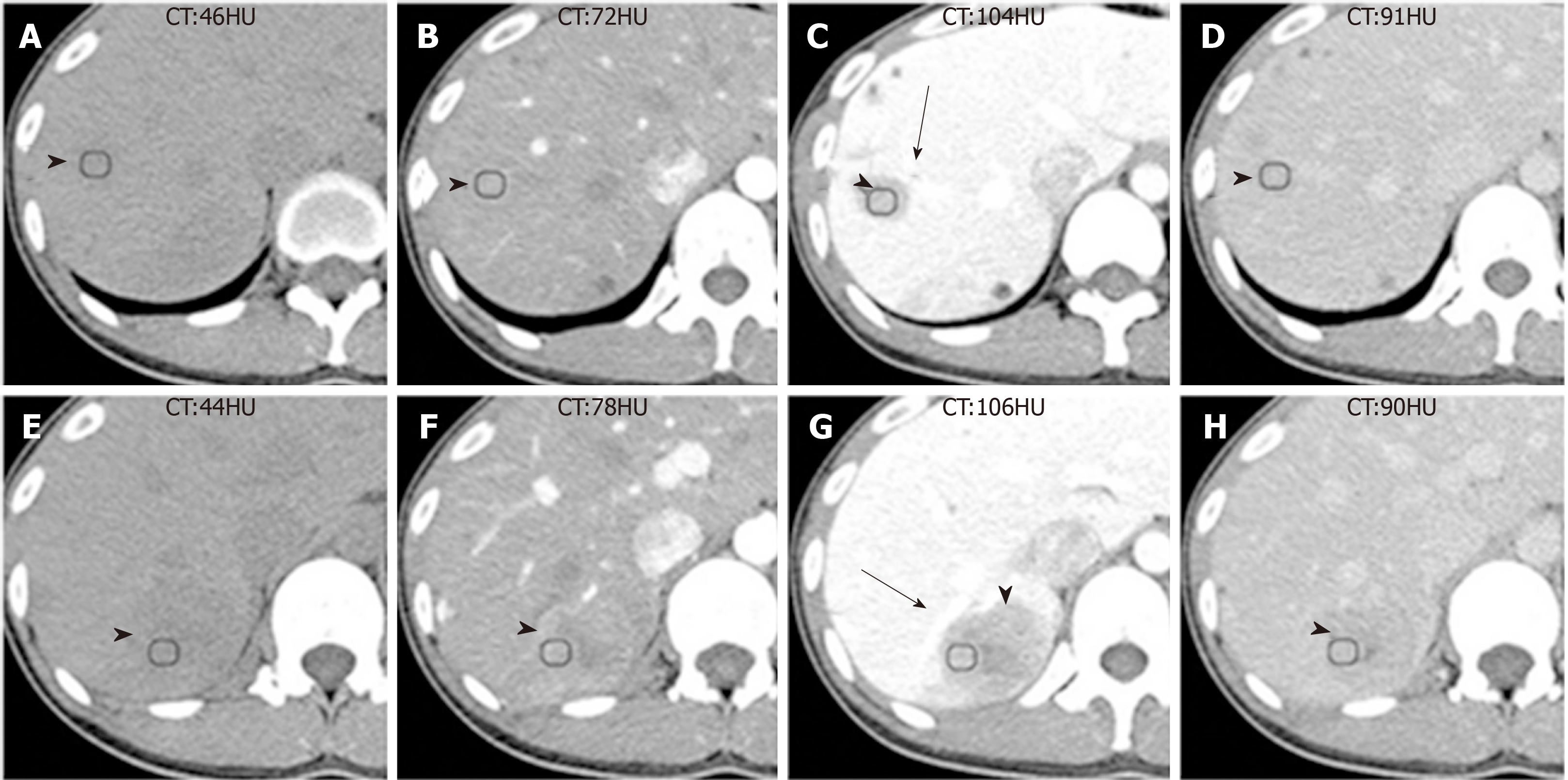
Figure 1 Computed tomography images of Case 1.
A and E: The unenhanced computed tomography (CT) images showed two well-defined hypodense masses; B-D, F-H: The triple-phase enhanced CT images showed both tumors had heterogeneous sustained hypoenhancement with internal necrosis. As the enhanced scan time was extended, the range of low density components within the mass decreased. Both tumors were close to the portal vein branch (black arrows) and always exhibited hypoattenuation relative to the surrounding liver parenchyma. Inflammatory pseudotumor-like follicular dendritic cell tumors (black arrowheads) from the upper segment of the right posterior lobe of the liver were present in a 31-year-old Chinese woman. CT: Computed tomography.
- Citation: Li HL, Liu HP, Guo GWJ, Chen ZH, Zhou FQ, Liu P, Liu JB, Wan R, Mao ZQ. Imaging findings of inflammatory pseudotumor-like follicular dendritic cell tumors of the liver: Two case reports and literature review. World J Gastroenterol 2019; 25(45): 6693-6703
- URL: https://www.wjgnet.com/1007-9327/full/v25/i45/6693.htm
- DOI: https://dx.doi.org/10.3748/wjg.v25.i45.6693


