Copyright
©The Author(s) 2019.
World J Gastroenterol. Dec 7, 2019; 25(45): 6634-6652
Published online Dec 7, 2019. doi: 10.3748/wjg.v25.i45.6634
Published online Dec 7, 2019. doi: 10.3748/wjg.v25.i45.6634
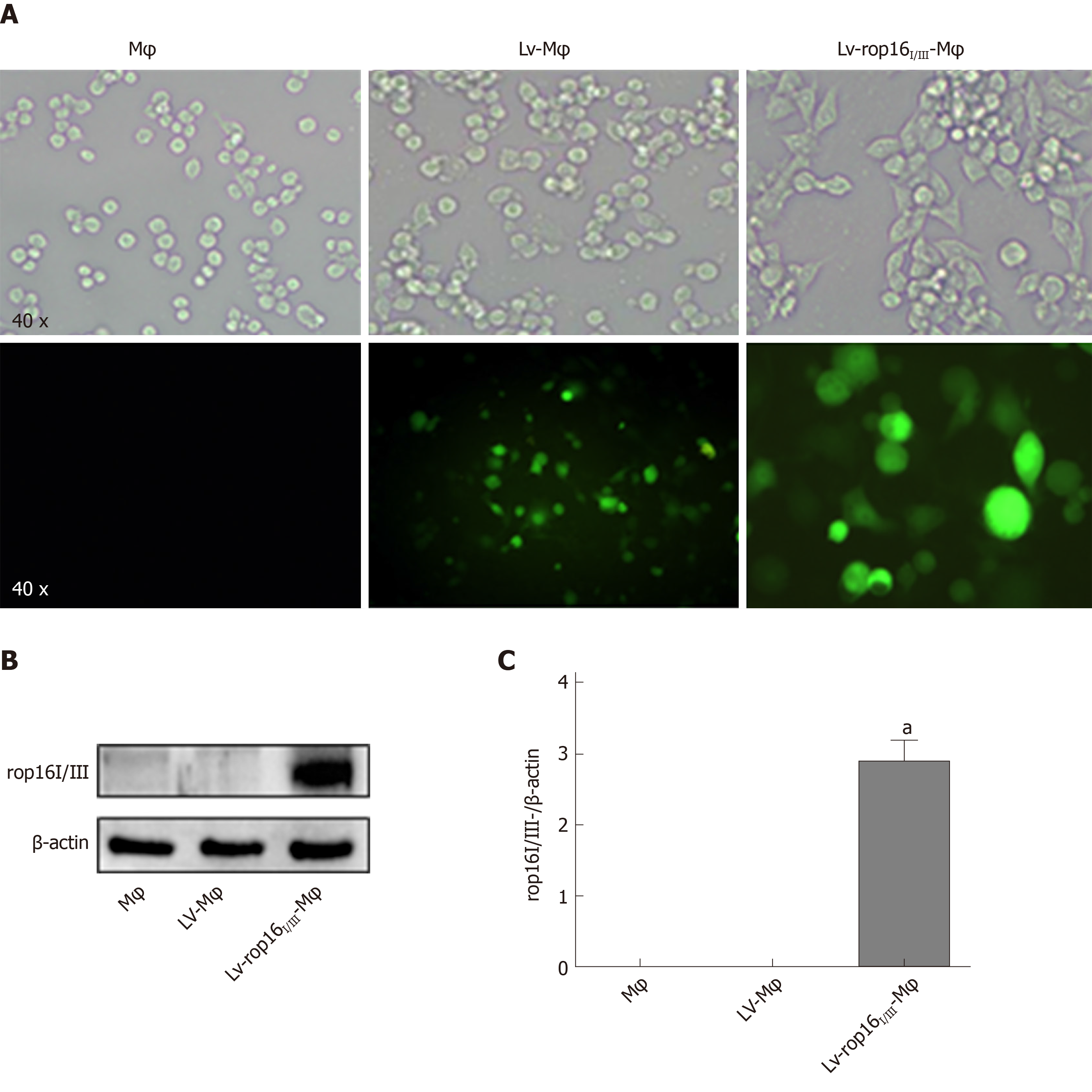
Figure 1 Stable transfection of RAW264.
7 cells with LV-rop16I/III recombinant lentivirus. A: Fluorescence microscopy was used to observe the expression of green fluorescent protein in macrophages, Lv-Mφ and Lv-rop16I/III-Mφ cells stably transfected with recombinant lentivirus. B: Macrophages, Lv-Mφ, and Lv-rop16I/III-Mφ stably-transfected cells were analyzed by Western blotting. C: Statistical analysis of protein expression in Lv-rop16I/III-Mφ cells relative to non-transfected macrophages and mock Lv-Mφ by Western blotting. aP < 0.001 vs Mφ. Mφ: Macrophages; LV-Mφ: Lentivirus transfer into macrophages; LV-rop16I/III-Mφ: Lentivirus-rop16I/III transfer into macrophages.
- Citation: Xu YW, Xing RX, Zhang WH, Li L, Wu Y, Hu J, Wang C, Luo QL, Shen JL, Chen X. Toxoplasma ROP16I/III ameliorated inflammatory bowel diseases via inducing M2 phenotype of macrophages. World J Gastroenterol 2019; 25(45): 6634-6652
- URL: https://www.wjgnet.com/1007-9327/full/v25/i45/6634.htm
- DOI: https://dx.doi.org/10.3748/wjg.v25.i45.6634


