Copyright
©The Author(s) 2019.
World J Gastroenterol. Nov 7, 2019; 25(41): 6190-6204
Published online Nov 7, 2019. doi: 10.3748/wjg.v25.i41.6190
Published online Nov 7, 2019. doi: 10.3748/wjg.v25.i41.6190
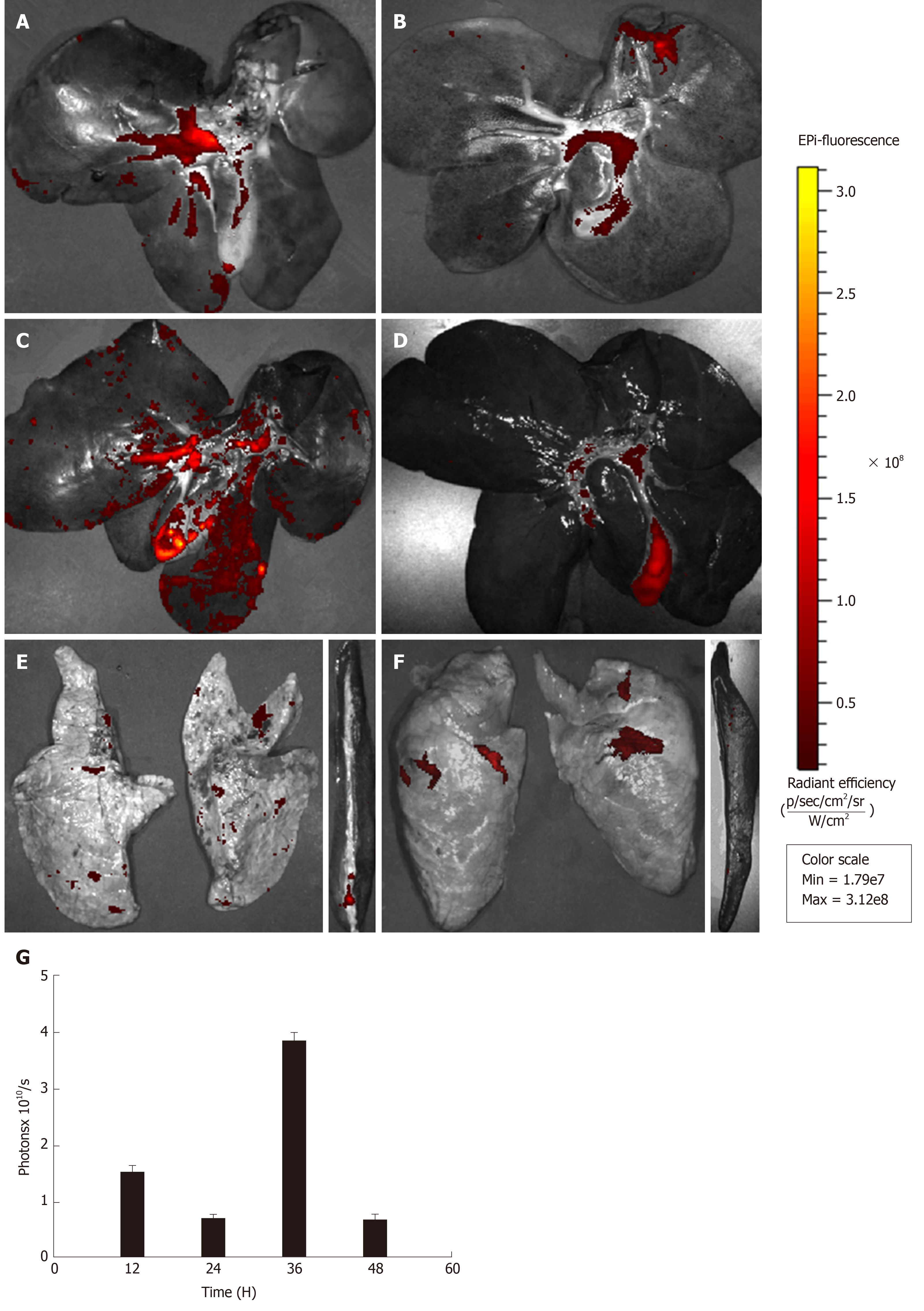
Figure 5 Ex vivo images showing the dynamic biodistribution of transplanted PKH26-labelled menstrual blood stem cells in different organs by the in vivo imaging system.
A: At 12 h after transplantation, strong red fluorescence emitted from PKH26-labelled menstrual blood stem cells (MenSCs) was detected in the liver; B: The fluorescence intensity of the liver weakened at 24 h; C: The fluorescence intensity of the liver increased notably at 36 h; D: Finally, the signal faded after 48 h; E: At 24 h after transplantation, a weak signal was detected in the lungs and the spleen; F: At 48 h after transplantation, a weak signal was also detected in the lungs, but it was seldom observed in the spleen; G: A graph showing the total photon emission efficiency in the liver at all time points. MenSCs: Menstrual blood stem cells; IVIS: In vivo Imaging System.
- Citation: Cen PP, Fan LX, Wang J, Chen JJ, Li LJ. Therapeutic potential of menstrual blood stem cells in treating acute liver failure. World J Gastroenterol 2019; 25(41): 6190-6204
- URL: https://www.wjgnet.com/1007-9327/full/v25/i41/6190.htm
- DOI: https://dx.doi.org/10.3748/wjg.v25.i41.6190


