Copyright
©The Author(s) 2019.
World J Gastroenterol. Nov 7, 2019; 25(41): 6190-6204
Published online Nov 7, 2019. doi: 10.3748/wjg.v25.i41.6190
Published online Nov 7, 2019. doi: 10.3748/wjg.v25.i41.6190
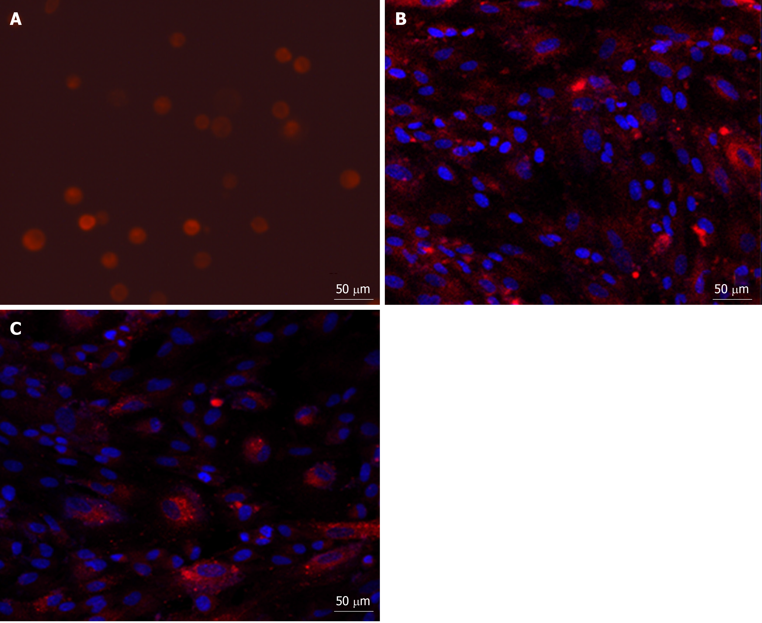
Figure 3 Fluorescence micrograph of PKH26-labelled menstrual blood stem cells.
A: The third passage of PKH26-labelled menstrual blood stem cells (MenSCs) was resuspended and exhibited red fluorescence (×20); B: When the labelled MenSCs were cultured in vitro, most of the cells were able to grow adhering to the wall (×20); C: After serial passages, the fluorescent signal intensity from the cells decreased with time (×20, passage 8). MenSCs: Menstrual blood stem cells.
- Citation: Cen PP, Fan LX, Wang J, Chen JJ, Li LJ. Therapeutic potential of menstrual blood stem cells in treating acute liver failure. World J Gastroenterol 2019; 25(41): 6190-6204
- URL: https://www.wjgnet.com/1007-9327/full/v25/i41/6190.htm
- DOI: https://dx.doi.org/10.3748/wjg.v25.i41.6190


