Copyright
©The Author(s) 2019.
World J Gastroenterol. Oct 28, 2019; 25(40): 6053-6062
Published online Oct 28, 2019. doi: 10.3748/wjg.v25.i40.6053
Published online Oct 28, 2019. doi: 10.3748/wjg.v25.i40.6053
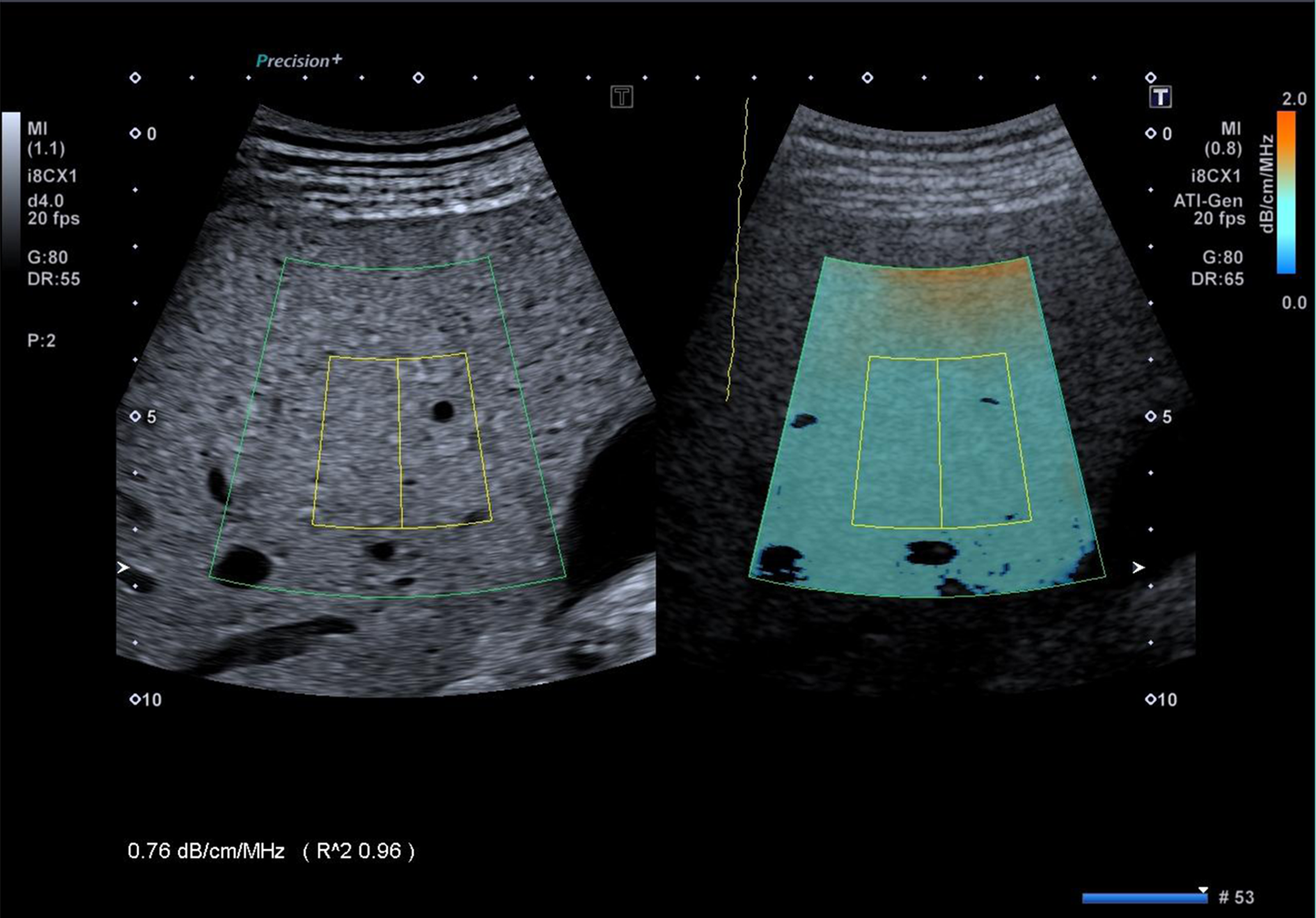
Figure 1 Intercostal scan of the right lobe of the liver, obtained with the Aplio i800 ultrasound system equipped with the ATI® technique.
The color-coded map, which is overlaid on the B-mode image, and the B-mode image without colors are shown side-by-side on the monitor of the ultrasound system. The inner rectangle is the fixed measurement box. The reliability of the result is displayed by the R2 value.
- Citation: Ferraioli G, Soares Monteiro LB. Ultrasound-based techniques for the diagnosis of liver steatosis. World J Gastroenterol 2019; 25(40): 6053-6062
- URL: https://www.wjgnet.com/1007-9327/full/v25/i40/6053.htm
- DOI: https://dx.doi.org/10.3748/wjg.v25.i40.6053


