Copyright
©The Author(s) 2019.
World J Gastroenterol. Jan 28, 2019; 25(4): 498-508
Published online Jan 28, 2019. doi: 10.3748/wjg.v25.i4.498
Published online Jan 28, 2019. doi: 10.3748/wjg.v25.i4.498
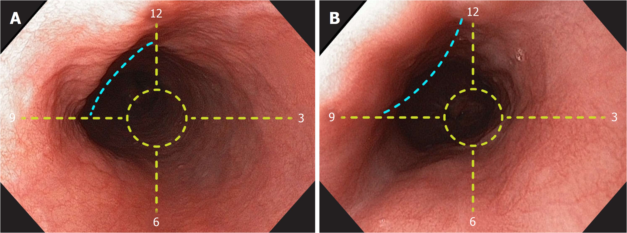
Figure 3 Identification of the left main bronchus and left atrium landmarks.
A: A concave, non-pulsatile, extrinsic esophageal compression (blue lines) is observed 25 cm from the incisors. The landmark is radially oriented at the anterior esophageal quadrant, between 9 and 12 o’clock. B: A convex, pulsatile, extrinsic esophageal compression (blue lines) is observed 31 cm from the incisors. The landmark is radially oriented at the anterior esophageal quadrant, between 9 and 12 o’clock.
- Citation: Emura F, Gomez-Esquivel R, Rodriguez-Reyes C, Benias P, Preciado J, Wallace M, Giraldo-Cadavid L. Endoscopic identification of endoluminal esophageal landmarks for radial and longitudinal orientation and lesion location. World J Gastroenterol 2019; 25(4): 498-508
- URL: https://www.wjgnet.com/1007-9327/full/v25/i4/498.htm
- DOI: https://dx.doi.org/10.3748/wjg.v25.i4.498


