Copyright
©The Author(s) 2019.
World J Gastroenterol. Sep 28, 2019; 25(36): 5569-5577
Published online Sep 28, 2019. doi: 10.3748/wjg.v25.i36.5569
Published online Sep 28, 2019. doi: 10.3748/wjg.v25.i36.5569
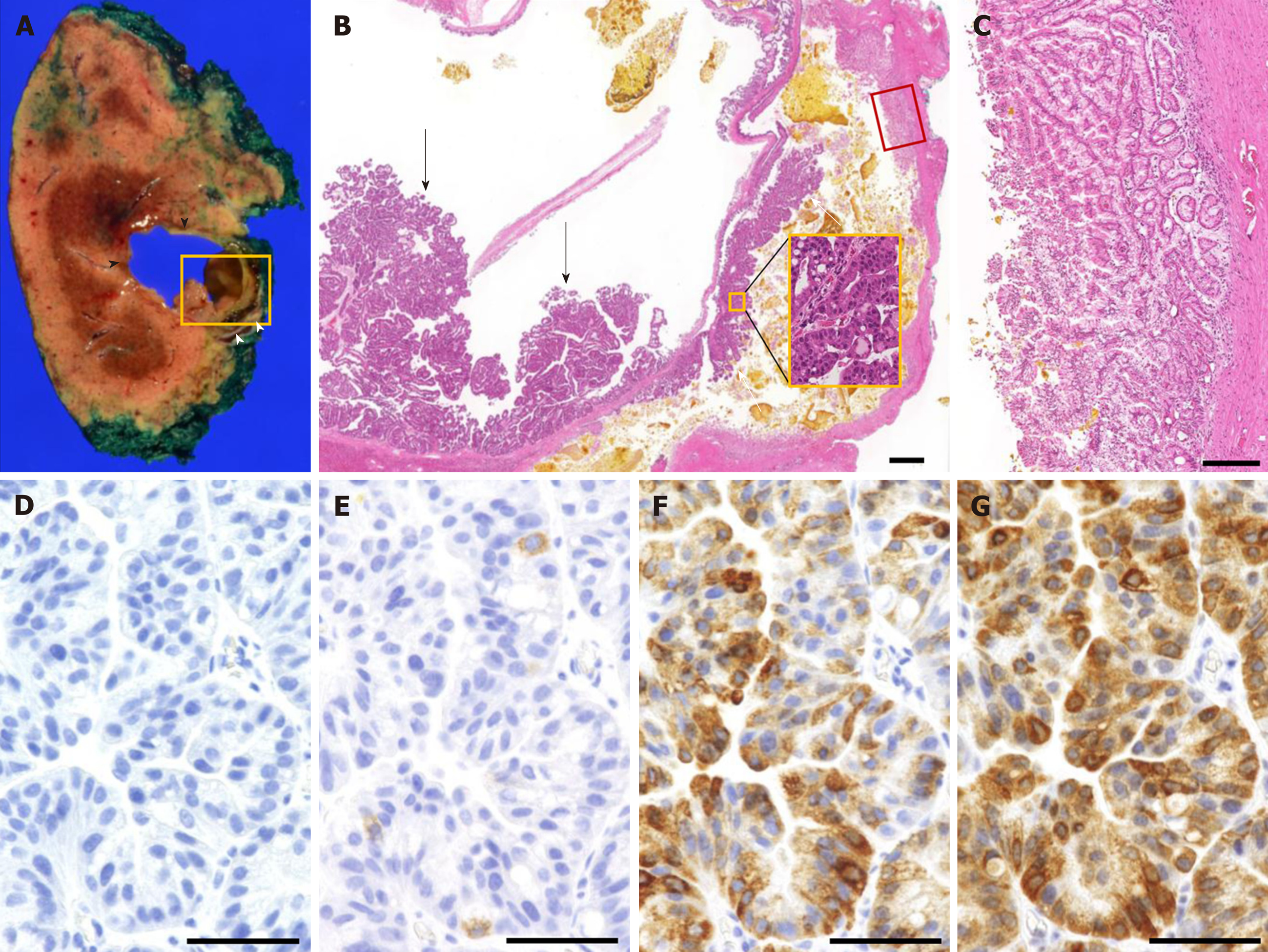
Figure 6 Pathological findings of the surgical specimens.
A: Macroscopic findings show 2 cystic lesions with a maximum diameter of 3 cm. The anterior tumor (black arrowhead) contained mucin and bile with papillary proliferation, while the posterior tumor (white arrowhead) contained solidified bile. B: Microscopic findings of the tumor (yellow square in A). Dysplastic epithelium proliferated on both anterior tumor (black arrow) and posterior tumor (white arrow). Higher magnification of the papillary proliferation of the posterior tumor (yellow square in B) shows dysplastic epithelium. C: Thick glandular epithelium was observed on the posterior tumor (red square in B), which corresponds to the hyperenhancement seen on the contrast-enhanced ultrasonography. D-G: Immunohistochemical findings of the papillary tumor show MUC1 negativity (D), MUC2 positivity in less than 5% of the cells (E), MUC5AC positivity (F), and MUC6 positivity (G). The black bar in B represents 1000 µm, C represents 200 µm, and D-G represents 50 µm. MUC: Mucin core protein.
- Citation: Hasebe T, Sawada K, Hayashi H, Nakajima S, Takahashi H, Hagiwara M, Imai K, Yuzawa S, Fujiya M, Furukawa H, Okumura T. Long-term growth of intrahepatic papillary neoplasms: A case report. World J Gastroenterol 2019; 25(36): 5569-5577
- URL: https://www.wjgnet.com/1007-9327/full/v25/i36/5569.htm
- DOI: https://dx.doi.org/10.3748/wjg.v25.i36.5569


