Copyright
©The Author(s) 2019.
World J Gastroenterol. Sep 7, 2019; 25(33): 4892-4903
Published online Sep 7, 2019. doi: 10.3748/wjg.v25.i33.4892
Published online Sep 7, 2019. doi: 10.3748/wjg.v25.i33.4892
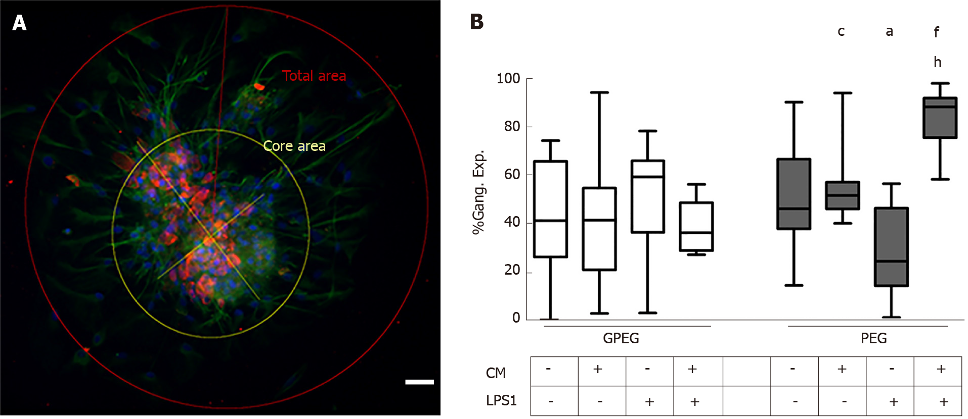
Figure 5 Effect of porcine vascular wall mesenchymal stromal cells supernatants on ganglion expansion.
A: Representative photo of the morphometric analysis performed to compare glial cells processes elongating from ganglion’s cores under different experimental conditions (scale bar: 100 µm); B: Relative area occupied by glial processes of guinea pig (left, gray bars) and pig enteric ganglia (right white bars). Guinea pig enteric ganglia did not show any significant difference between the different treatment groups. Conversely, pig enteric ganglia were more subjected to morphological changes: There was a decrease of the expanded area in the group treated with lipopolysaccharide 1 µg/mL (LPS1) compared to control (-42.88%, aP < 0.05). Moreover, conditioned medium (CM) derived by porcine vascular wall mesenchymal stromal cells evoked a higher protrusion of glial processes than LPS1 alone (+36.8%, cP < 0.05) which was remarkably higher in combination with LPS1 (CM+LPS1, +43.2% vs CTRL, fP < 0.01; +60.9% vs LPS1, hP < 0.01). LPS1: Lipopolysaccharide 1 µg/mL; CM: Conditioned medium; GPEG: Guinea pig enteric ganglia; PEG: Pig enteric ganglia.
- Citation: Dothel G, Bernardini C, Zannoni A, Spirito MR, Salaroli R, Bacci ML, Forni M, Ponti FD. Ex vivo effect of vascular wall stromal cells secretome on enteric ganglia. World J Gastroenterol 2019; 25(33): 4892-4903
- URL: https://www.wjgnet.com/1007-9327/full/v25/i33/4892.htm
- DOI: https://dx.doi.org/10.3748/wjg.v25.i33.4892


