
















©The Author(s) 2019.
World J Gastroenterol. Aug 14, 2019; 25(30): 4213-4221
Published online Aug 14, 2019. doi: 10.3748/wjg.v25.i30.4213
Published online Aug 14, 2019. doi: 10.3748/wjg.v25.i30.4213
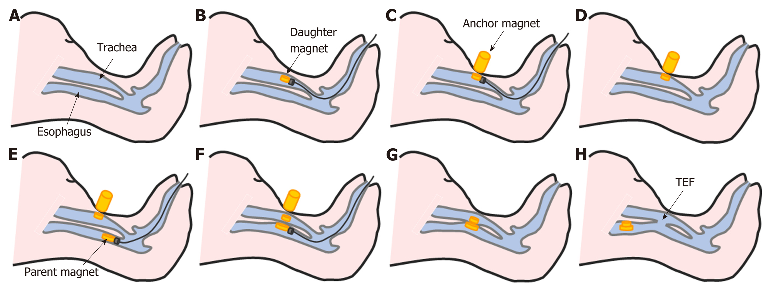
Figure 2 Study procedures.
A: Normal anatomy of the trachea and esophagus in the beagle; B: The daughter magnet was inserted into the trachea and advanced to the target location using the transmission magnet and the guide wire; C: The anchor magnet was placed on the neck of the animal over the trachea to fix the daughter magnet in its target location; D: The guide wire and transmission magnet were withdrawn from the trachea; E: The parent magnet was inserted into the cervical esophagus and advanced to the target location using the transmission magnet and the guide wire; F: The parent magnet and daughter magnet were attracted to each other by magnetic force; G: The guide wire and transmission magnet were withdrawn from the esophagus, and the anchor magnet was removed; H: 4-6 d later, the necrotic tissue between the two magnets had been shed and the parent magnet and daughter magnet had detached from the target location. The tracheoesophageal fistula was formed.
- Citation: Gao Y, Wu RQ, Lv Y, Yan XP. Novel magnetic compression technique for establishment of a canine model of tracheoesophageal fistula. World J Gastroenterol 2019; 25(30): 4213-4221
- URL: https://www.wjgnet.com/1007-9327/full/v25/i30/4213.htm
- DOI: https://dx.doi.org/10.3748/wjg.v25.i30.4213