Copyright
©The Author(s) 2019.
World J Gastroenterol. Jul 28, 2019; 25(28): 3753-3763
Published online Jul 28, 2019. doi: 10.3748/wjg.v25.i28.3753
Published online Jul 28, 2019. doi: 10.3748/wjg.v25.i28.3753
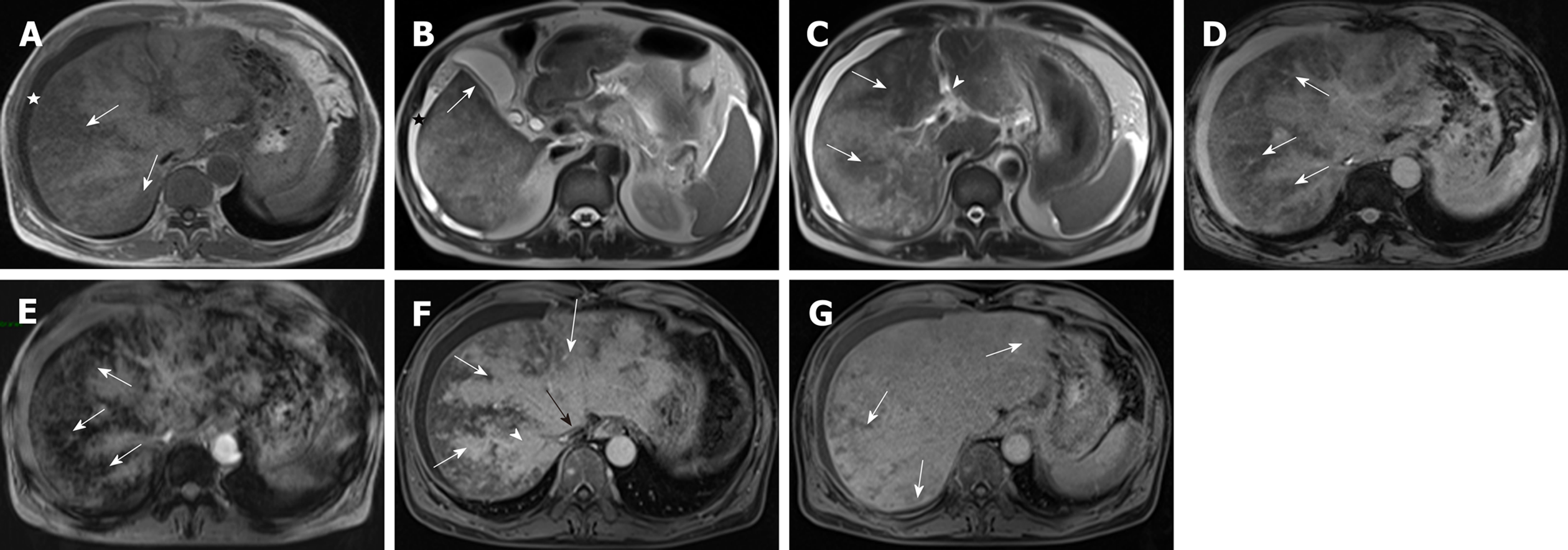
Figure 3 A 53-year-old man with pyrrolizidine alkaloids-induced hepatic sinusoidal obstruction syndrome (same patient) received gadoxetic acid-enhanced magnetic resonance imaging scan.
A: T1-weighted imaging. Ascites (white star), heterogeneous hypointensity (white arrow) were shown; B and C: T2-weighted imaging. Imaging findings included ascites (black star, B), gallbladder wall thickening (white arrow, B), periportal edema (arrowhead, C), heterogeneous hypointensity (white arrow, C); D and E: T2*WI (D) and SWI (E). Heterogeneous hypointensity (white arrow) was observed, and distribution of hypointensity in SWI and T2*WI was similar; F: Image of portal venous phase. Patchy liver enhancement, “claw-shaped” enhancement surrounding hepatic veins (white arrow), stenosis of right hepatic vein (arrowhead) and inferior vena cava (black arrow); G: Image of hepatobiliary phase. Heterogeneous hypointensity (white arrow) was shown. SWI: Susceptibility-weighted imaging; T2*WI: T2*-weighted imaging.
- Citation: Yang XQ, Ye J, Li X, Li Q, Song YH. Pyrrolizidine alkaloids-induced hepatic sinusoidal obstruction syndrome: Pathogenesis, clinical manifestations, diagnosis, treatment, and outcomes. World J Gastroenterol 2019; 25(28): 3753-3763
- URL: https://www.wjgnet.com/1007-9327/full/v25/i28/3753.htm
- DOI: https://dx.doi.org/10.3748/wjg.v25.i28.3753


