Copyright
©The Author(s) 2019.
World J Gastroenterol. Jan 14, 2019; 25(2): 269-281
Published online Jan 14, 2019. doi: 10.3748/wjg.v25.i2.269
Published online Jan 14, 2019. doi: 10.3748/wjg.v25.i2.269
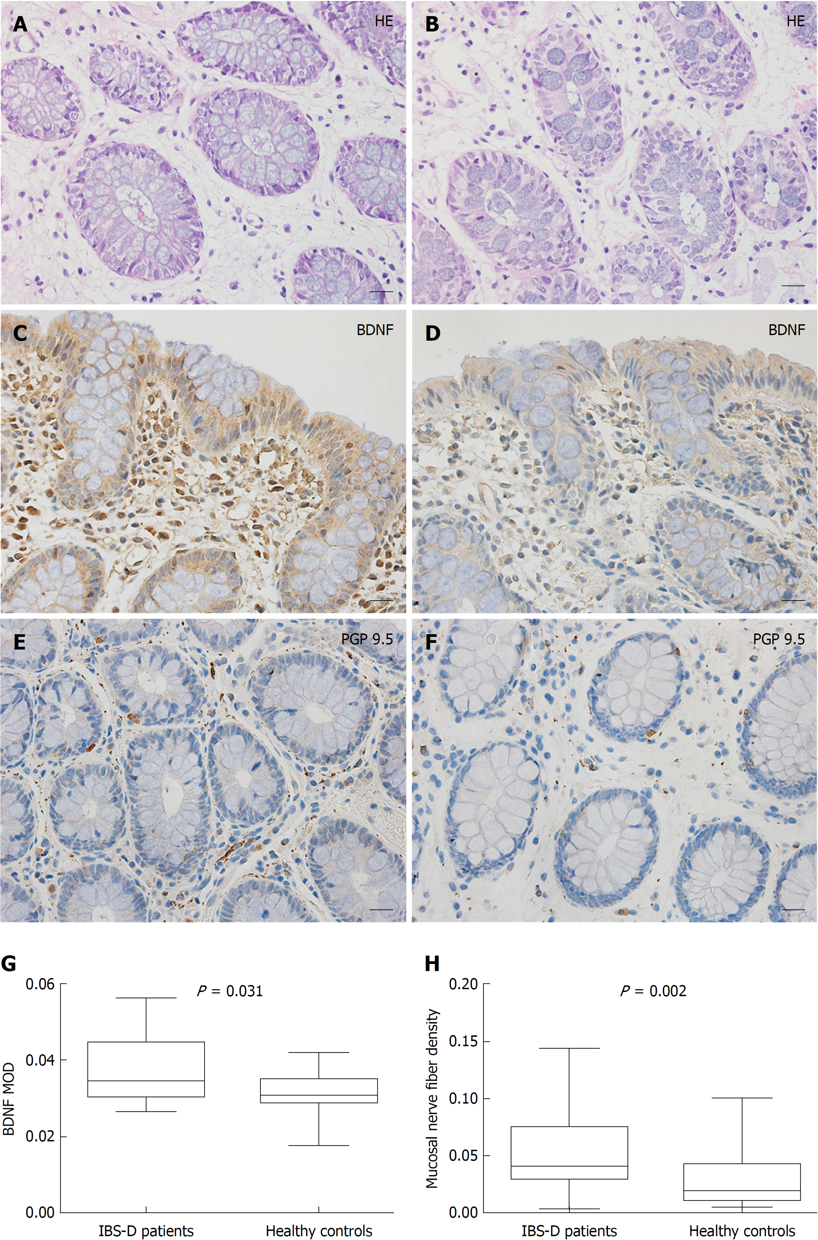
Figure 2 Mucosal histology and immunohistochemistry in patients with diarrhea-predominant irritable bowel syndrome and healthy controls.
A and B: Mucosae of diarrhea-predominant irritable bowel syndrome (IBS-D) patients and healthy controls both exhibited a normal histology (Scale bar = 20 μm); C and D: Brain-derived neurotrophic factor (BDNF) immunoreactivity was mainly scattered in the epithelium and lamina propria in IBS-D patients (Scale bar = 20 μm); E and F: Protein gene product (PGP) 9.5-immunoreactive nerve fibers were distributed in the lamina propria, showing fibrous appearance. (Scale bar = 20 μm). G: Mean optical density of BDNF in the colonic mucosa in IBS-D patients was significantly higher than that in controls (P = 0.031); H: The area occupied by PGP 9.5-immunoreactive fibers was greater in patients than in controls (P = 0.002). IBS-D: Diarrhea-predominant irritable bowel syndrome; BDNF: Brain-derived neurotrophic factor; H-E: Haematoxylin and eosin; PGP: Protein gene product; MOD: Mean optical density.
- Citation: Zhang Y, Qin G, Liu DR, Wang Y, Yao SK. Increased expression of brain-derived neurotrophic factor is correlated with visceral hypersensitivity in patients with diarrhea-predominant irritable bowel syndrome. World J Gastroenterol 2019; 25(2): 269-281
- URL: https://www.wjgnet.com/1007-9327/full/v25/i2/269.htm
- DOI: https://dx.doi.org/10.3748/wjg.v25.i2.269


