Copyright
©The Author(s) 2019.
World J Gastroenterol. May 14, 2019; 25(18): 2264-2270
Published online May 14, 2019. doi: 10.3748/wjg.v25.i18.2264
Published online May 14, 2019. doi: 10.3748/wjg.v25.i18.2264
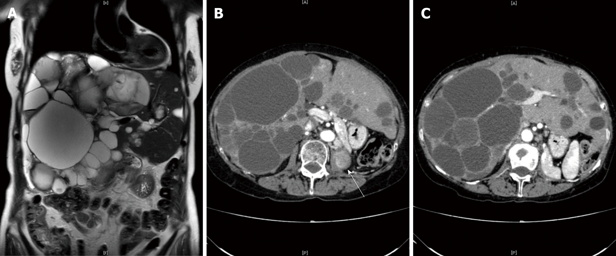
Figure 2 Images from abdominal computed tomography.
Local recurrence of renal cell carcinoma was detected in the ipsilateral lymph nodes and in the left lumbar vertebra (B, white arrow). There was a diffuse involvement of the liver parenchyma, with multiple cysts and large areas of noncystic liver parenchyma remaining (C); A: coronal view; B and C: transverse view.
- Citation: Liang C, Takahashi K, Kurata M, Sakashita S, Oda T, Ohkohchi N. Recurrent renal cell carcinoma leading to a misdiagnosis of polycystic liver disease: A case report. World J Gastroenterol 2019; 25(18): 2264-2270
- URL: https://www.wjgnet.com/1007-9327/full/v25/i18/2264.htm
- DOI: https://dx.doi.org/10.3748/wjg.v25.i18.2264


