Copyright
©The Author(s) 2019.
World J Gastroenterol. May 7, 2019; 25(17): 2144-2148
Published online May 7, 2019. doi: 10.3748/wjg.v25.i17.2144
Published online May 7, 2019. doi: 10.3748/wjg.v25.i17.2144
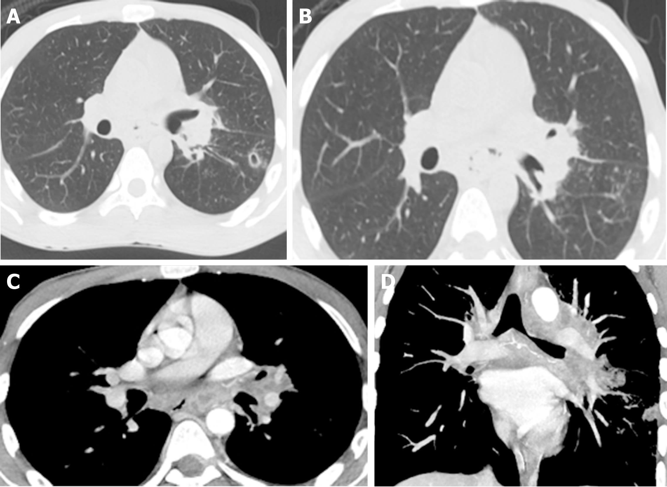
Figure 2 Computed tomography chest.
A: Computed tomography lung window axial image shows left lower lobe lung cavitary lesion with multiple nodules in tree-in-bud configuration, a classical sign of lung tuberculosis; B: Chest computed tomography lung window axial image shows multiple mediastinal air pockets anterior to the esophagus, indicating an esophageal fistula; C: Computed tomography chest with IV contrast mediastinal window axial image shows multiple mediastinal necrotic lymph nodes and mediastinal air pocket anterior to esophagus indicating mediastinal tuberculosis and esophageal fistula; D: Computed tomography chest with IV contrast maximum intensity projection coronal reformatted image shows mediastinal bronchial artery aneurysm.
- Citation: Alharbi SR. Tuberculous esophagomediastinal fistula with concomitant mediastinal bronchial artery aneurysm-acute upper gastrointestinal bleeding: A case report. World J Gastroenterol 2019; 25(17): 2144-2148
- URL: https://www.wjgnet.com/1007-9327/full/v25/i17/2144.htm
- DOI: https://dx.doi.org/10.3748/wjg.v25.i17.2144


