Copyright
©The Author(s) 2019.
World J Gastroenterol. Apr 28, 2019; 25(16): 2010-2018
Published online Apr 28, 2019. doi: 10.3748/wjg.v25.i16.2010
Published online Apr 28, 2019. doi: 10.3748/wjg.v25.i16.2010
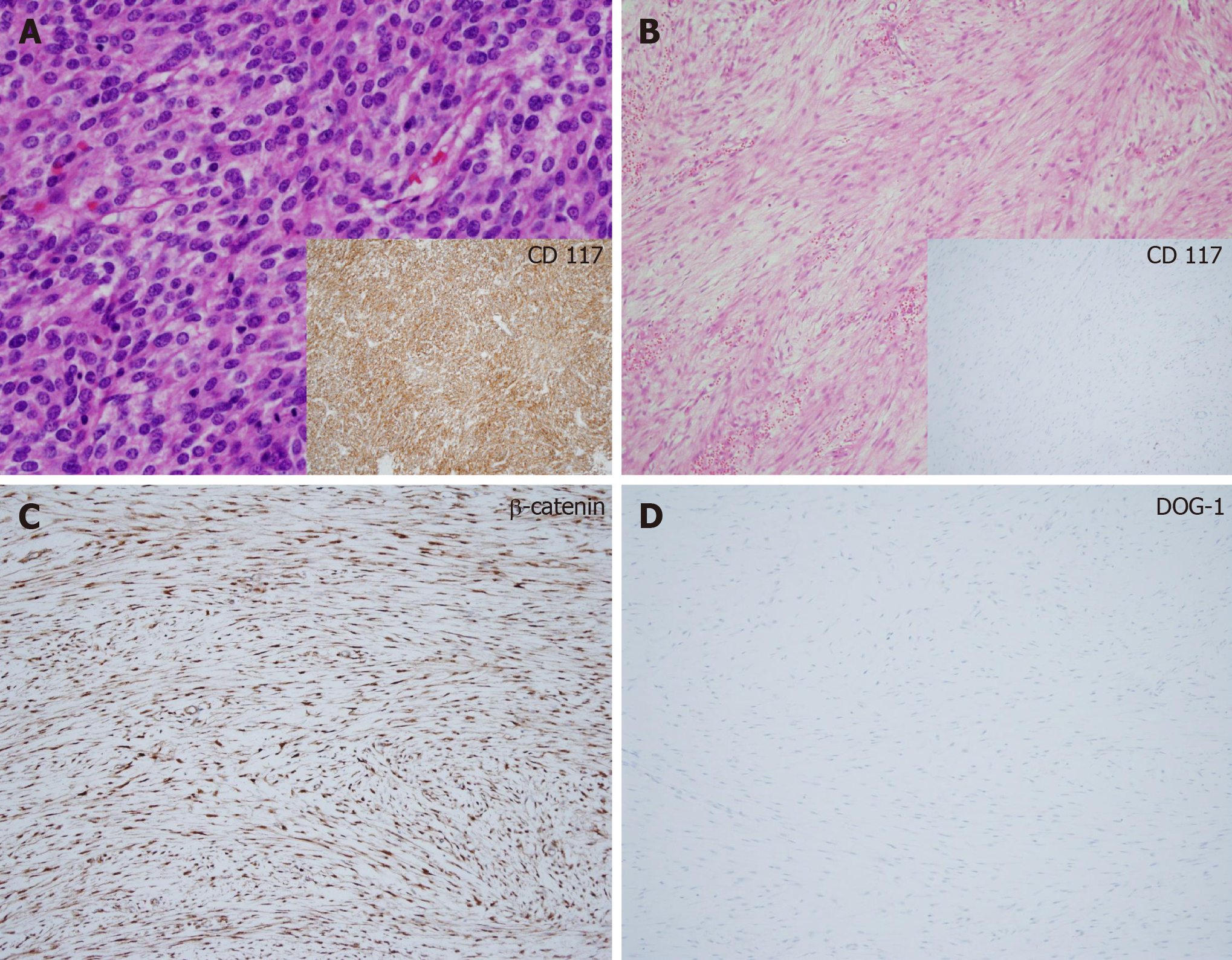
Figure 2 Histological examination of intra-abdominal desmoid tumors in patients with gastrointestinal stromal tumors showed low or moderate cellularity with proliferative spindle cells in a fibrotic background and infiltrative growth patterns (hematoxylin and eosin, original magnification × 400).
Tumor cells were negative for CD117 and DOG-1, whereas positive for β-catenin. A: Patient 7: A 40-year-old female patient presented with small bowel gastrointestinal stromal tumors (GIST) with metastasis in the peritoneum, liver, and ovary. She underwent initial debulking surgery. GIST cells were positive for CD117; B: Desmoid tumor (DT) in the pelvic peritoneum showed low cellularity with collagen fibers and was negative for CD117; C: DT was positive for β-catenin; D: DT was negative for DOG-1. GIST: Gastrointestinal stromal tumors; DT: Desmoid tumor.
- Citation: Kim JH, Ryu MH, Park YS, Kim HJ, Park H, Kang YK. Intra-abdominal desmoid tumors mimicking gastrointestinal stromal tumors — 8 cases: A case report. World J Gastroenterol 2019; 25(16): 2010-2018
- URL: https://www.wjgnet.com/1007-9327/full/v25/i16/2010.htm
- DOI: https://dx.doi.org/10.3748/wjg.v25.i16.2010


