Copyright
©The Author(s) 2019.
World J Gastroenterol. Apr 28, 2019; 25(16): 2010-2018
Published online Apr 28, 2019. doi: 10.3748/wjg.v25.i16.2010
Published online Apr 28, 2019. doi: 10.3748/wjg.v25.i16.2010
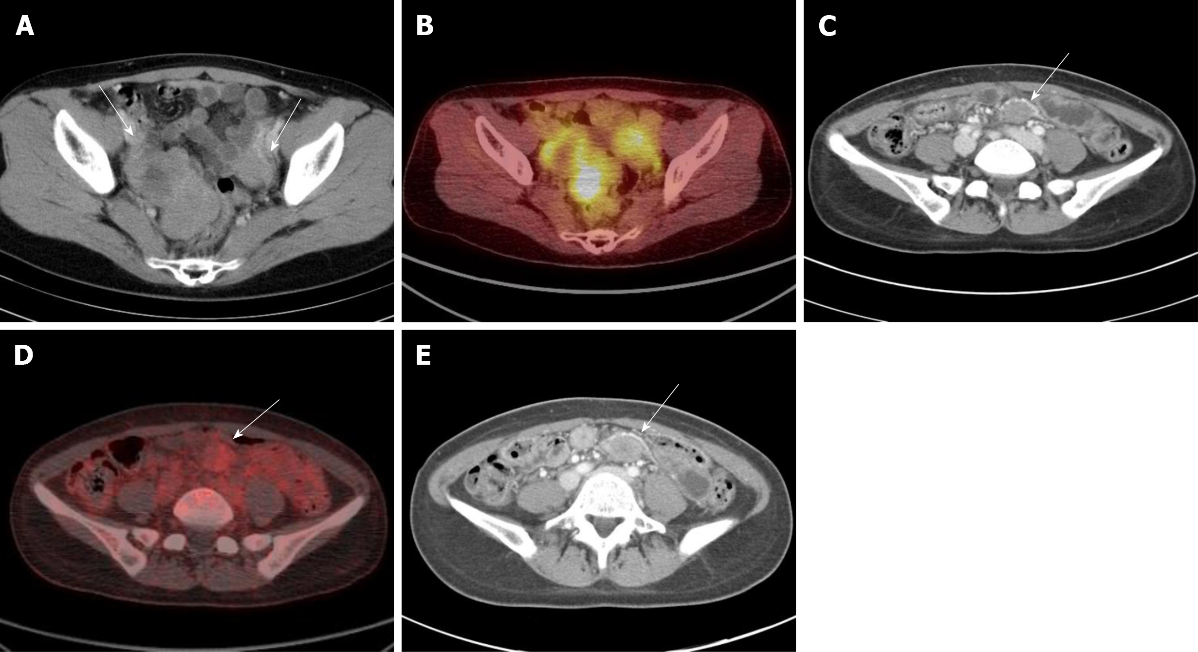
Figure 1 Abdominal computed tomography and 18fluorodeoxyglucose-positron emission tomography revealed intra-abdominal desmoid tumors in patients with gastrointestinal stromal tumors, showing well-defined ovoid shaped masses with delayed or mild enhancement on computed tomography and mild or absence of hypermetabolic activity on 18fluorodeoxyglucose-positron emission tomography.
A: Patient 7: A 40-year-old female patient presented with small bowel gastrointestinal stromal tumors (GIST) with metastasis in the peritoneum, liver, and ovary. Pelvic lobulated masses with heterogeneous enhancement were observed on computed tomography; B: Initial GIST lesions showed hypermetabolic activity on 18fluorodeoxyglucose-positron emission tomography (18FDG-PET); C: A new single mass occurred in the pelvic peritoneum; D: The maximum standardized uptake values was 2.7 as shown on 18FDG-PET; E: The dose of imatinib was escalated to 800 mg/d and the size of desmoid tumors was stable for approximately 1.3 years. 18FDG-PET: 18Fluorodeoxyglucose-positron emission tomography; GIST: Gastrointestinal stromal tumors.
- Citation: Kim JH, Ryu MH, Park YS, Kim HJ, Park H, Kang YK. Intra-abdominal desmoid tumors mimicking gastrointestinal stromal tumors — 8 cases: A case report. World J Gastroenterol 2019; 25(16): 2010-2018
- URL: https://www.wjgnet.com/1007-9327/full/v25/i16/2010.htm
- DOI: https://dx.doi.org/10.3748/wjg.v25.i16.2010


