Copyright
©The Author(s) 2019.
World J Gastroenterol. Apr 7, 2019; 25(13): 1531-1549
Published online Apr 7, 2019. doi: 10.3748/wjg.v25.i13.1531
Published online Apr 7, 2019. doi: 10.3748/wjg.v25.i13.1531
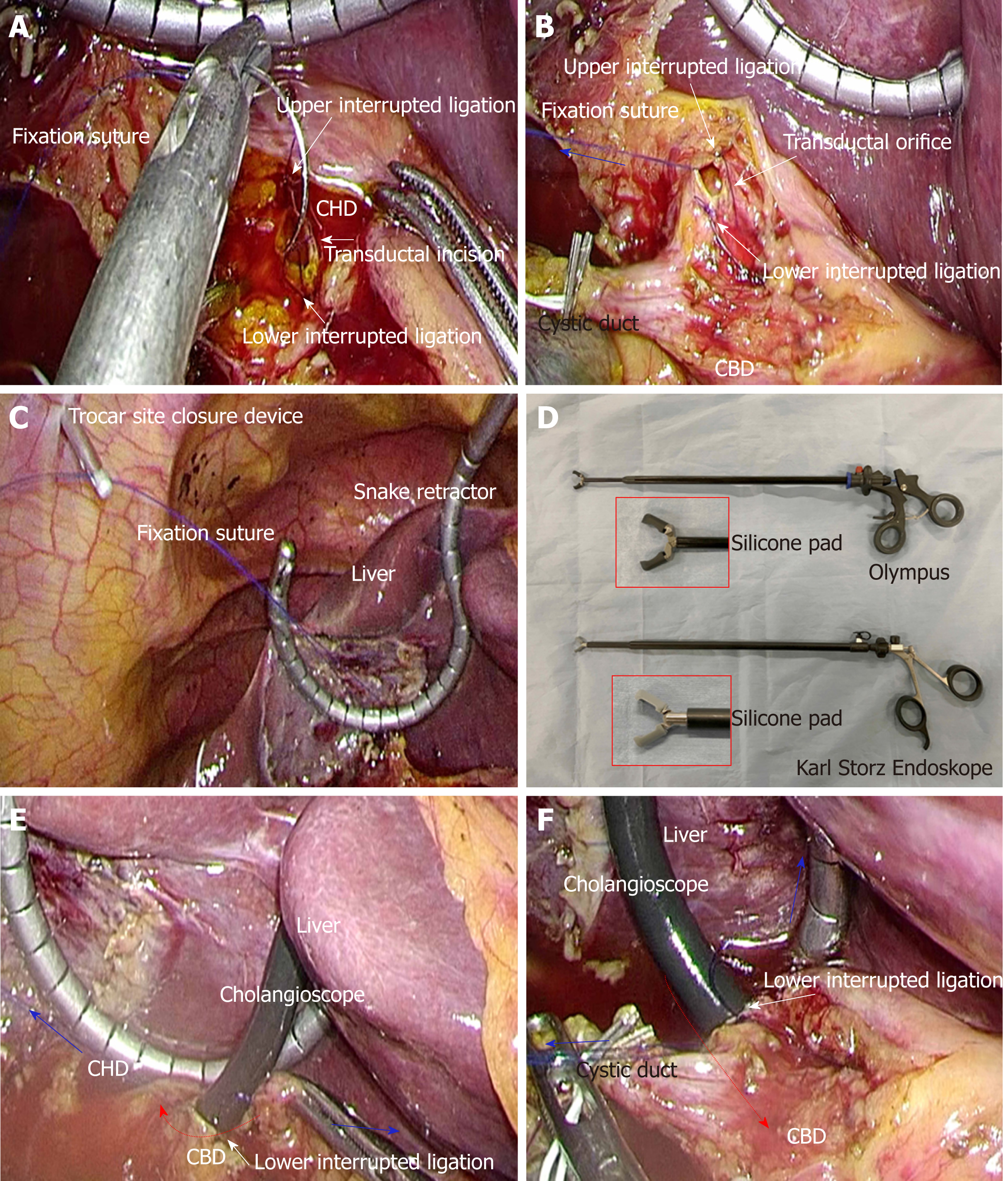
Figure 5 Actual surgical procedures of laparoscopic choledocholithotomy.
A and B: Fixation sutures (blue arrow) are bilaterally placed to open the transductal orifice; C: Fixation sutures are adequately set through the abdominal wall at different points from the laparoscopic trocars with a trocar site closure device. The liver is held cranially with a snake retractor to stretch the hepatoduodenal ligament; D: A dedicated elastic forceps is important for successful laparoscopic choledocholithotomy. The tip of the forceps contains a silicone pad to avoid damaging the cholangioscope. Olympus (A66070A; Tokyo, Japan) and Karl Storz Endoskope (K33531 PG; Tuttlingen, Germany) provide made-to-order forceps, respectively; E and F: The bifurcation of hepatic ducts on the common hepatic duct side (E) and characteristic findings of the end of the intra-pancreatic bile duct on the common bile duct side (F) should be confirmed. Interrupted ligations at the upper and lower edges of the transductal incision prevent progressive laceration during cholangioscope maneuvers (red arrows). Fixation sutures (blue arrows) are removed. CHD: Common hepatic duct; CBD: Common bile duct.
- Citation: Hori T. Comprehensive and innovative techniques for laparoscopic choledocholithotomy: A surgical guide to successfully accomplish this advanced manipulation. World J Gastroenterol 2019; 25(13): 1531-1549
- URL: https://www.wjgnet.com/1007-9327/full/v25/i13/1531.htm
- DOI: https://dx.doi.org/10.3748/wjg.v25.i13.1531


