Copyright
©The Author(s) 2019.
World J Gastroenterol. Mar 28, 2019; 25(12): 1465-1477
Published online Mar 28, 2019. doi: 10.3748/wjg.v25.i12.1465
Published online Mar 28, 2019. doi: 10.3748/wjg.v25.i12.1465
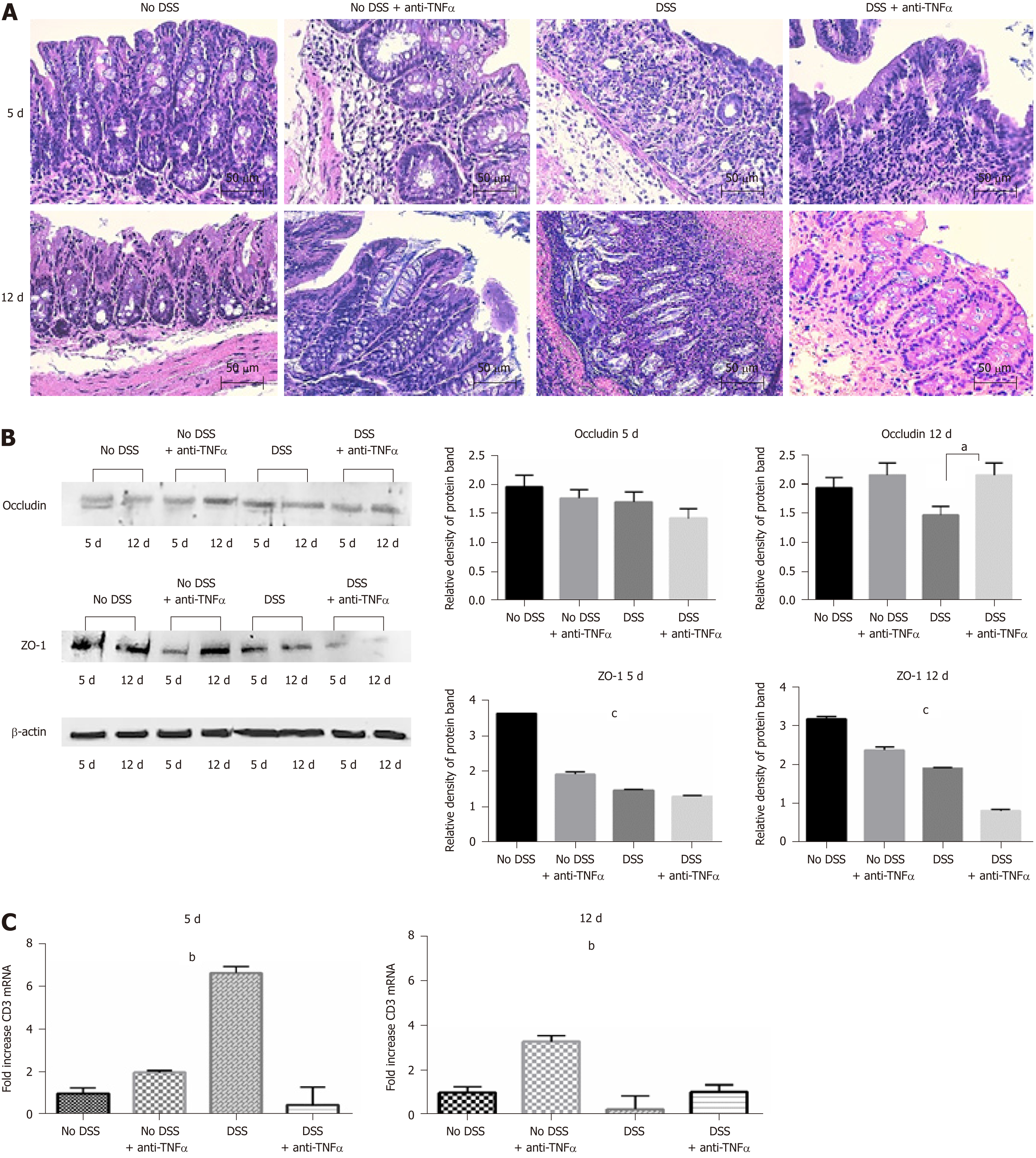
Figure 3 Histological analysis confirms the strength of the dextran sulfate sodium model.
A: Hematoxylin and eosin staining of colon sections of mice from 4 groups of treatment at day 5 and 12. Forty-fold magnification was used. B: Protein expression of tight junctions occludin and ZO-1 in colon tissue. Antibody anti-β-actin was used as endogenous control. On the right, the histograms represent the mean of densitometry values from 3 different experiments. C: CD3 infiltration in colon mucosa was assessed by quantitative PCR of CD3 mRNA. One-way ANOVA (and non-parametric) was calculated; aP < 0.05; bP < 0.01; cP < 0.0001. DSS: Dextran sulfate sodium; TNFα: Tumor necrosis factor α.
- Citation: Petito V, Graziani C, Lopetuso LR, Fossati M, Battaglia A, Arena V, Scannone D, Quaranta G, Quagliariello A, Del Chierico F, Putignani L, Masucci L, Sanguinetti M, Sgambato A, Gasbarrini A, Scaldaferri F. Anti-tumor necrosis factor α therapy associates to type 17 helper T lymphocytes immunological shift and significant microbial changes in dextran sodium sulphate colitis. World J Gastroenterol 2019; 25(12): 1465-1477
- URL: https://www.wjgnet.com/1007-9327/full/v25/i12/1465.htm
- DOI: https://dx.doi.org/10.3748/wjg.v25.i12.1465


