Copyright
©The Author(s) 2019.
World J Gastroenterol. Mar 14, 2019; 25(10): 1259-1265
Published online Mar 14, 2019. doi: 10.3748/wjg.v25.i10.1259
Published online Mar 14, 2019. doi: 10.3748/wjg.v25.i10.1259
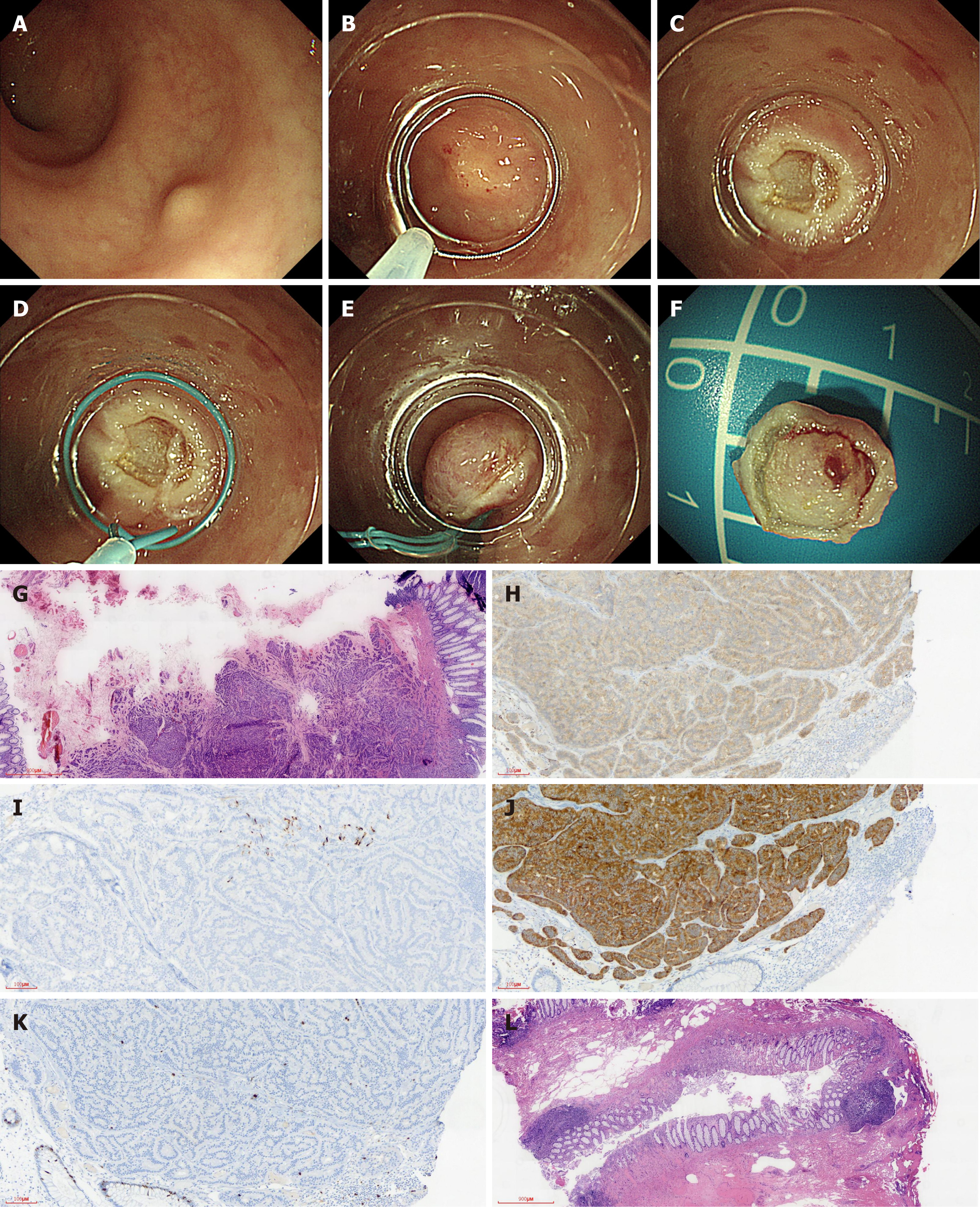
Figure 1 Utilizing endoloop ligation after cap-endoscopic mucosal resection using a transparent cap to remove rectal carcinoid tumor.
Pathology suggested positive margins and further transanal endoscopic microsurgery pathology was negative. A: Endoscopy showing a rectal carcinoid about 10 mm in diameter; B: An electric snare mounted on the transparent cap on the inner lens end; C: Wound after resection; D: Nylon endoloop device installed on the inner lens end; E: Wound after nylon endoloop ligation resection; F: Endoscopic resection of the intact tumor; G: Pathological specimen suggesting a carcinoid, vertical margin positive (× 10); H: Positive immunohistochemical staining for CD56 (× 100); I: Positive immunohistochemical staining for chromogrin (× 100); J: Positive immunohistochemical staining for Syn (× 100); K: Immunohistochemical staining for Ki-67 (< 2%; × 100); L: Transanal endoscopic microsurgery surgery did not identify tumor cells (× 10).
- Citation: Zhang DG, Luo S, Xiong F, Xu ZL, Li YX, Yao J, Wang LS. Endoloop ligation after endoscopic mucosal resection using a transparent cap: A novel method to treat small rectal carcinoid tumors. World J Gastroenterol 2019; 25(10): 1259-1265
- URL: https://www.wjgnet.com/1007-9327/full/v25/i10/1259.htm
- DOI: https://dx.doi.org/10.3748/wjg.v25.i10.1259


