Copyright
©The Author(s) 2019.
World J Gastroenterol. Mar 14, 2019; 25(10): 1197-1209
Published online Mar 14, 2019. doi: 10.3748/wjg.v25.i10.1197
Published online Mar 14, 2019. doi: 10.3748/wjg.v25.i10.1197
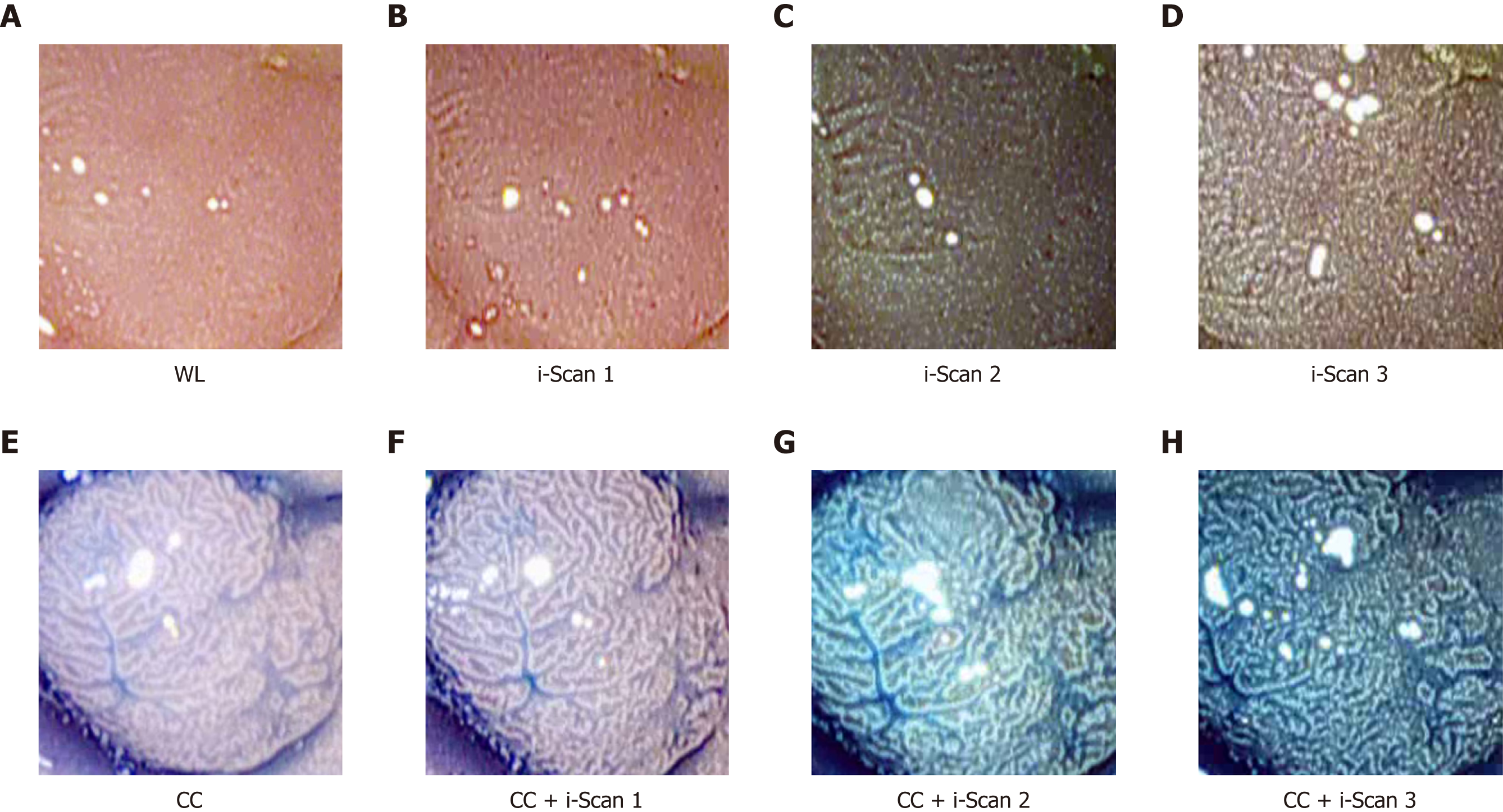
Figure 1 Images of a polyp using digital (i-Scan) and/or conventional chromoendoscopy.
WL: White light endoscopy; CC: Conventional chromoendoscopy.
- Citation: Wimmer G, Gadermayr M, Wolkersdörfer G, Kwitt R, Tamaki T, Tischendorf J, Häfner M, Yoshida S, Tanaka S, Merhof D, Uhl A. Quest for the best endoscopic imaging modality for computer-assisted colonic polyp staging. World J Gastroenterol 2019; 25(10): 1197-1209
- URL: https://www.wjgnet.com/1007-9327/full/v25/i10/1197.htm
- DOI: https://dx.doi.org/10.3748/wjg.v25.i10.1197


