Copyright
©The Author(s) 2018.
World J Gastroenterol. Feb 21, 2018; 24(7): 870-875
Published online Feb 21, 2018. doi: 10.3748/wjg.v24.i7.870
Published online Feb 21, 2018. doi: 10.3748/wjg.v24.i7.870
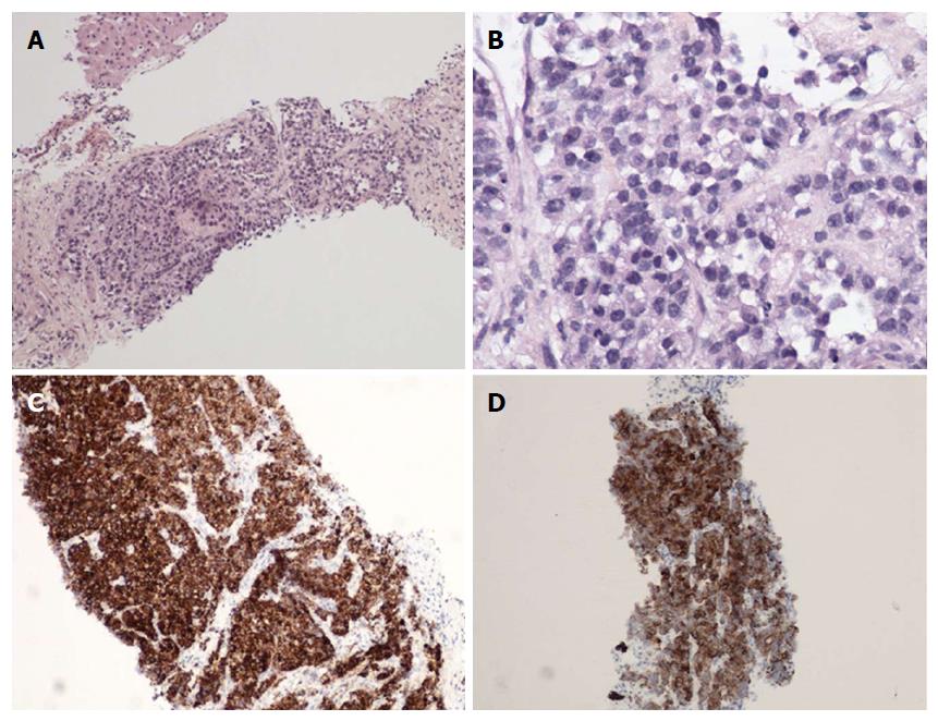
Figure 3 Histological and immunohistochemical appearance of hepatic lesions.
A: Combined hepatocellular cholangiocarcinoma, stem cell features, intermediate cell-subtype with underlying cirrhosis; B: The tumor is composed of small tumor cells arranged in bays, with some ill-defined glands; C: Cells express both markers of hepatocyte cells (Her Par1); D: Markers of biliary cells (cytokeratin 19).
- Citation: Salimon M, Chapelle N, Matysiak-Budnik T, Mosnier JF, Frampas E, Touchefeu Y. Esophageal metastasis of stem cell-subtype hepatocholangiocarcinoma: Rare presentation of a rare tumor. World J Gastroenterol 2018; 24(7): 870-875
- URL: https://www.wjgnet.com/1007-9327/full/v24/i7/870.htm
- DOI: https://dx.doi.org/10.3748/wjg.v24.i7.870


