Copyright
©The Author(s) 2018.
World J Gastroenterol. Feb 21, 2018; 24(7): 870-875
Published online Feb 21, 2018. doi: 10.3748/wjg.v24.i7.870
Published online Feb 21, 2018. doi: 10.3748/wjg.v24.i7.870
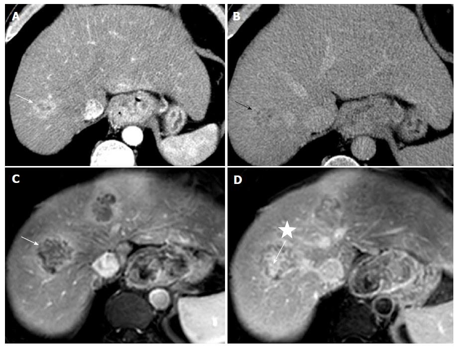
Figure 2 Computed tomography scan and magnetic resonance imaging scan imaging.
Axial-enhanced computed tomography scans with arterial (A) and 5-min delayed times (B). Corresponding axial enhanced MRI in T1-weighted sequence with fat suppression (C and D). Tumor of the junction of the segments VIII-VII combined the double imaging features with peripheral arterial contrast enhancement (white arrow) and secondary wash out (black arrow) (HCC part) and a late fibrous contrast enhancement of the central part (white asterisk) (ICC part). MRI was performed at 2-mo intervals and demonstrated a second tumor with comparable behavior in segment IV. CT: Computed tomography; MRI: Magnetic resonance imaging.
- Citation: Salimon M, Chapelle N, Matysiak-Budnik T, Mosnier JF, Frampas E, Touchefeu Y. Esophageal metastasis of stem cell-subtype hepatocholangiocarcinoma: Rare presentation of a rare tumor. World J Gastroenterol 2018; 24(7): 870-875
- URL: https://www.wjgnet.com/1007-9327/full/v24/i7/870.htm
- DOI: https://dx.doi.org/10.3748/wjg.v24.i7.870


