Copyright
©The Author(s) 2018.
World J Gastroenterol. Dec 28, 2018; 24(48): 5491-5504
Published online Dec 28, 2018. doi: 10.3748/wjg.v24.i48.5491
Published online Dec 28, 2018. doi: 10.3748/wjg.v24.i48.5491
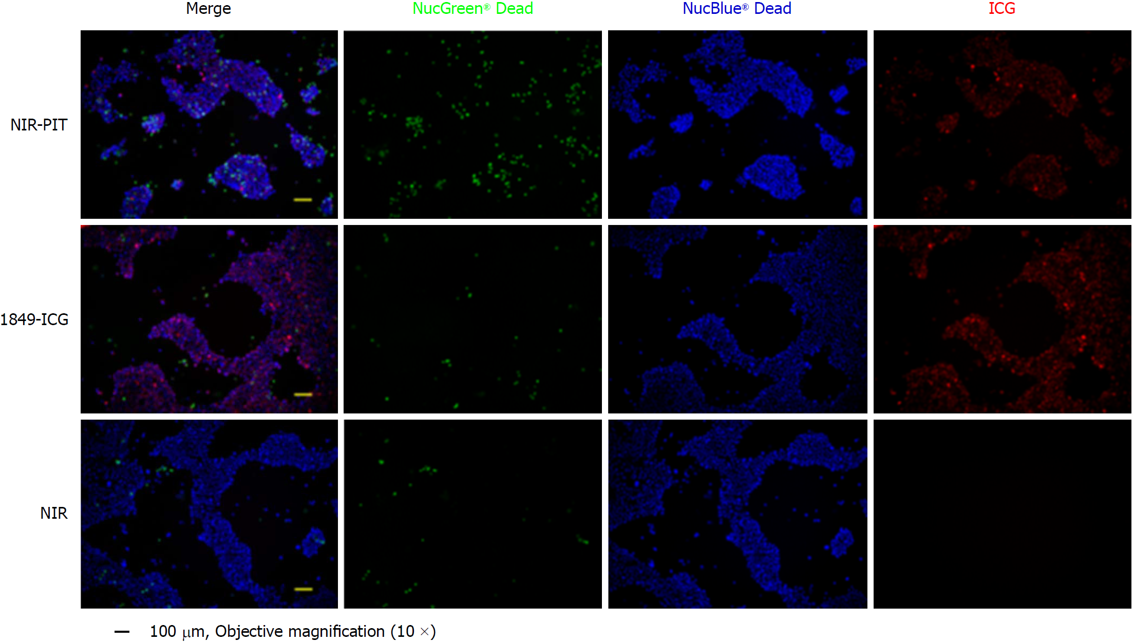
Figure 3 Phototoxic cell death after photoimmunotherapy.
Two hours after photoimmunotherapy (PIT) treatment, the cells were stained using the Cell viability imaging kit. The nuclei of dead cells were stained with NucGreen® Dead reagent (green), while the viable cell nuclei were stained with NucBlue® Live reagent (blue). Near-infrared (NIR)-PIT induced rapid cell death and numerous dead cells were markedly visible. There was no significant cytotoxicity associated with exposure to indocyanine green-labeled anti-tissue factor antibody 1849 or NIR light only. (Scale bar = 100 μm, 10 × objective magnification). 1849-ICG: Indocyanine green-labeled anti-tissue factor antibody 1849; NIR: Near-infrared; PIT: Photoimmunotherapy.
- Citation: Aung W, Tsuji AB, Sugyo A, Takashima H, Yasunaga M, Matsumura Y, Higashi T. Near-infrared photoimmunotherapy of pancreatic cancer using an indocyanine green-labeled anti-tissue factor antibody. World J Gastroenterol 2018; 24(48): 5491-5504
- URL: https://www.wjgnet.com/1007-9327/full/v24/i48/5491.htm
- DOI: https://dx.doi.org/10.3748/wjg.v24.i48.5491


