Copyright
©The Author(s) 2018.
World J Gastroenterol. Dec 21, 2018; 24(47): 5366-5378
Published online Dec 21, 2018. doi: 10.3748/wjg.v24.i47.5366
Published online Dec 21, 2018. doi: 10.3748/wjg.v24.i47.5366
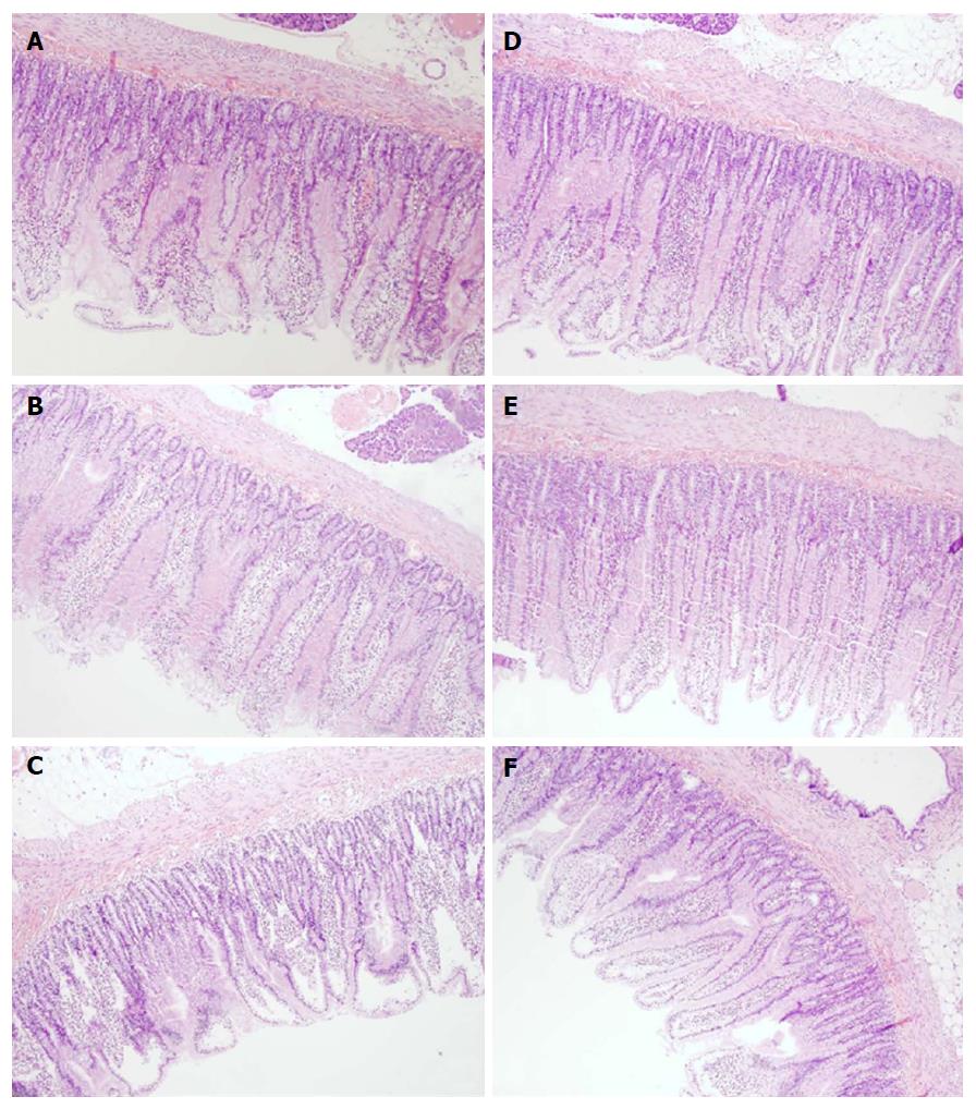
Figure 7 Microscopical presentation of duodenal lesions.
Microscopically (Hex10), in controls the lesions progressed from mild villous edema with mild lymphocytic infiltrate (5 min ligation time) (A) toward denuded villous tops with marked villous edema and submucosal capillary congestion (30 min ligation time) (B) to the substantial subepithelial space with abundant lifting of epithelial layer from lamina propria extending down sides of villi, villous edema with capillary congestion, submucosal congestion and lymphocytic infiltrate (24 h ligation time) (C). BPC 157 rats (D, E, F) exhibited always intestinal preservation with only mild villous edema and mild lymphocytic infiltrate. Elevation of epithelium from lamina propria was found only on the apical portion of villi (24 h ligation time, F).
- Citation: Amic F, Drmic D, Bilic Z, Krezic I, Zizek H, Peklic M, Klicek R, Pajtak A, Amic E, Vidovic T, Rakic M, Milkovic Perisa M, Horvat Pavlov K, Kokot A, Tvrdeic A, Boban Blagaic A, Zovak M, Seiwerth S, Sikiric P. Bypassing major venous occlusion and duodenal lesions in rats, and therapy with the stable gastric pentadecapeptide BPC 157, L-NAME and L-arginine. World J Gastroenterol 2018; 24(47): 5366-5378
- URL: https://www.wjgnet.com/1007-9327/full/v24/i47/5366.htm
- DOI: https://dx.doi.org/10.3748/wjg.v24.i47.5366


