Copyright
©The Author(s) 2018.
World J Gastroenterol. Dec 14, 2018; 24(46): 5215-5222
Published online Dec 14, 2018. doi: 10.3748/wjg.v24.i46.5215
Published online Dec 14, 2018. doi: 10.3748/wjg.v24.i46.5215
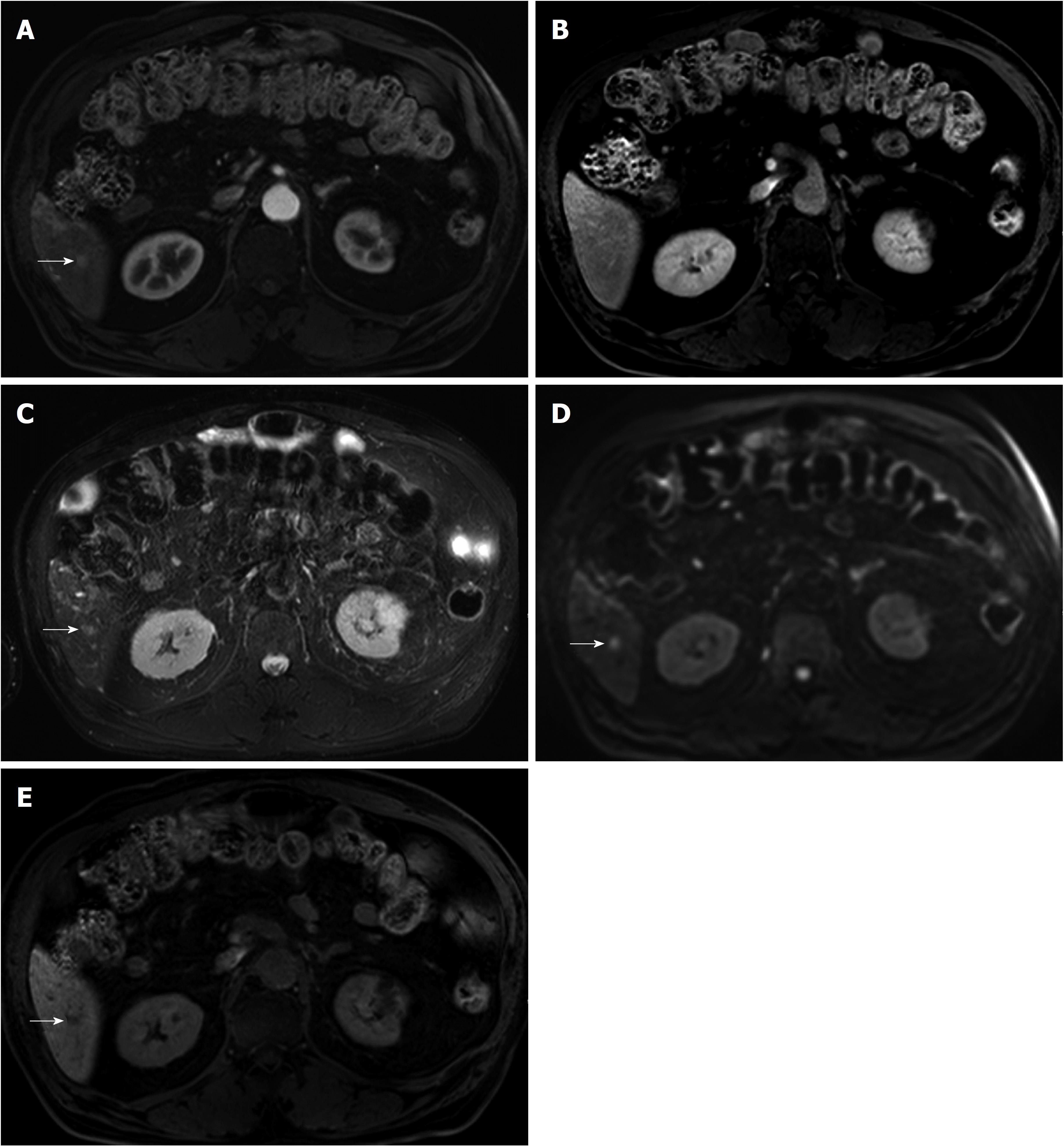
Figure 2 78-year-old man with a history of radiofrequency ablation for hepatocellular carcinoma.
A 6-mm enhancing nodule in segment 6 of the liver is seen on magnetic resonance imaging (MRI). Although the nodular lesion (arrow) shows arterial enhancement (A), it did not show washout on transitional phase (B). The lesion shows high signal intensity on both T2- and diffusion-weighted images (C and D), and low signal intensity on hepatobiliary phase (E). As the lesion did not show typical imaging features of hepatocellular carcinoma (HCC), the patient was followed with MRI. At follow up MRI obtained 3 mo later, the lesion disappeared (now shown here), suggesting the lesion was not HCC.
- Citation: Lee MW, Lim HK. Management of sub-centimeter recurrent hepatocellular carcinoma after curative treatment: Current status and future. World J Gastroenterol 2018; 24(46): 5215-5222
- URL: https://www.wjgnet.com/1007-9327/full/v24/i46/5215.htm
- DOI: https://dx.doi.org/10.3748/wjg.v24.i46.5215


