Copyright
©The Author(s) 2018.
World J Gastroenterol. Nov 28, 2018; 24(44): 5013-5024
Published online Nov 28, 2018. doi: 10.3748/wjg.v24.i44.5013
Published online Nov 28, 2018. doi: 10.3748/wjg.v24.i44.5013
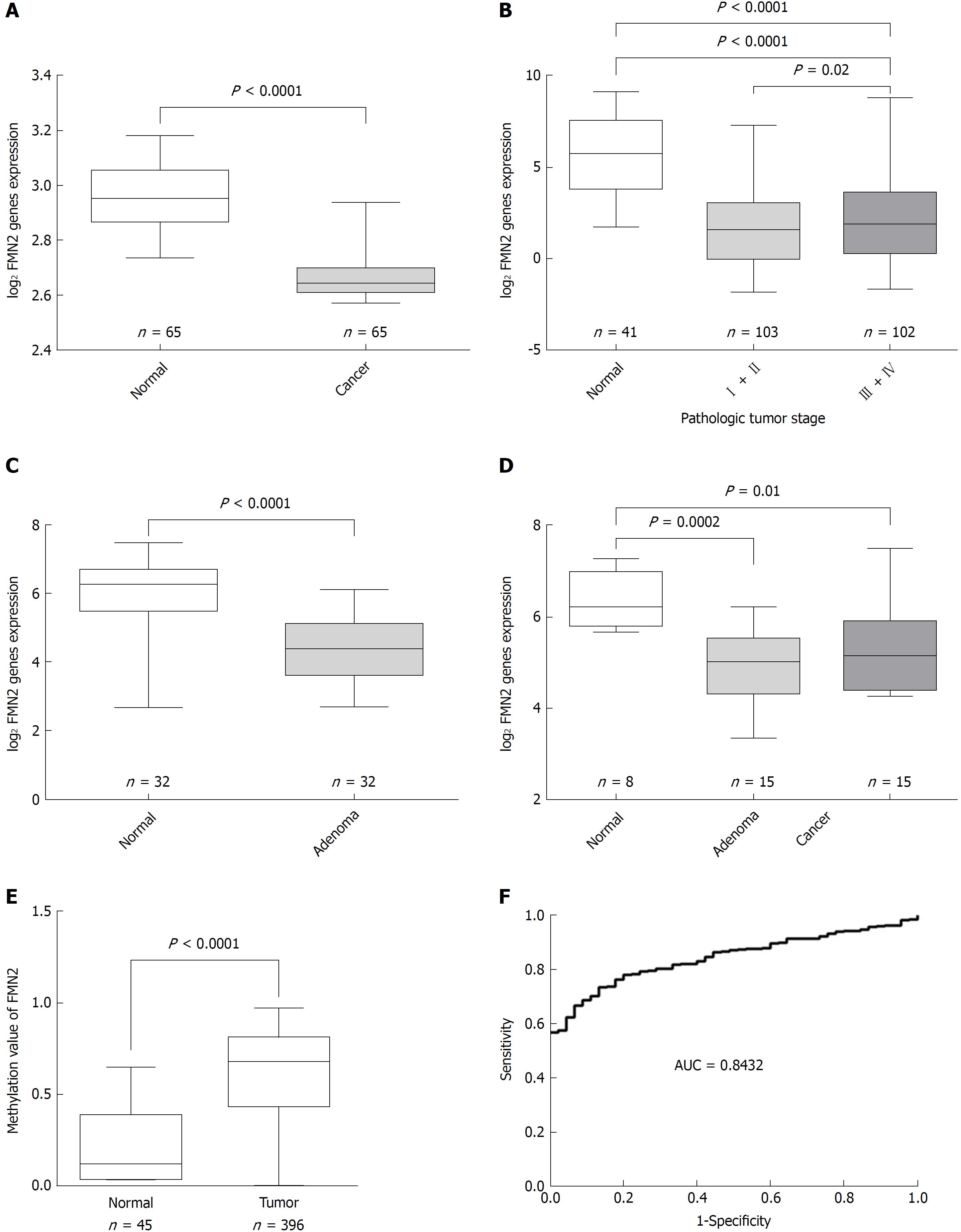
Figure 1 Colorectal tissues show decreased formin 2 expression and a high frequency of hypermethylation in the formin 2 promoter.
A: Normalized expression of formin 2 (FMN2) mRNA in carcinoma and adjacent normal tissues, presented as box-whisker plots (unpaired t-test, GEO: GSE20842); B: Normalized expression of FMN2 mRNA in tissue from different stages of carcinoma (according to the AJCC Cancer Staging Manual) and in normal tissue, presented as box-whisker plots (unpaired t-test, colorectal cancer samples from TCGA); C: Normalized expression of FMN2 mRNA in adenoma and corresponding normal colonic mucosal tissues, presented as box-whisker plots (unpaired t-test, GEO: GSE8671); D: Normalized expression of FMN2 mRNA in normal, adenoma, and carcinoma tissues, presented as box-whisker plots (unpaired t-test, GEO: GSE4183); E: The FMN2 gene shows an increased methylation level in colorectal cancer tissues compared with normal tissues (unpaired t-test, colorectal cancer samples from TCGA); F: Receiver operating characteristic curve analysis was used to assess the clinical diagnostic utility of FMN2 DNA methylation for the prediction of colorectal cancer.
- Citation: Li DJ, Feng ZC, Li XR, Hu G. Involvement of methylation-associated silencing of formin 2 in colorectal carcinogenesis. World J Gastroenterol 2018; 24(44): 5013-5024
- URL: https://www.wjgnet.com/1007-9327/full/v24/i44/5013.htm
- DOI: https://dx.doi.org/10.3748/wjg.v24.i44.5013


