Copyright
©The Author(s) 2018.
World J Gastroenterol. Jan 28, 2018; 24(4): 537-542
Published online Jan 28, 2018. doi: 10.3748/wjg.v24.i4.537
Published online Jan 28, 2018. doi: 10.3748/wjg.v24.i4.537
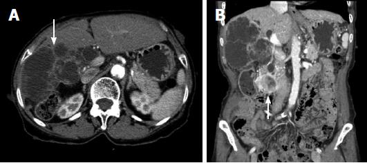
Figure 2 Computed tomography.
A: A low-attenuated multilocular mass with septation, and a mural nodule containing a soft tissue-enhancing lesion in the right hepatic lobe (white arrow); B: Diffuse intrahepatic duct (IHD) dilatation, especially in the right hepatic lobe, with a suspicious soft tissue lesion connected to the IHD. A 1.7-cm-sized, well-demarcated enhancing mass is observed in the third portion of the duodenum (white arrow). 300 mm × 225 mm (300 × 300 DPI).
- Citation: Lee JM, Lee JM, Hyun JJ, Choi HS, Kim ES, Keum B, Jeen YT, Chun HJ, Lee HS, Kim CD, Kim DS, Kim JY. Intraductal papillary bile duct adenocarcinoma and gastrointestinal stromal tumor in a case of neurofibromatosis type 1. World J Gastroenterol 2018; 24(4): 537-542
- URL: https://www.wjgnet.com/1007-9327/full/v24/i4/537.htm
- DOI: https://dx.doi.org/10.3748/wjg.v24.i4.537


