Copyright
©The Author(s) 2018.
World J Gastroenterol. Oct 14, 2018; 24(38): 4356-4368
Published online Oct 14, 2018. doi: 10.3748/wjg.v24.i38.4356
Published online Oct 14, 2018. doi: 10.3748/wjg.v24.i38.4356
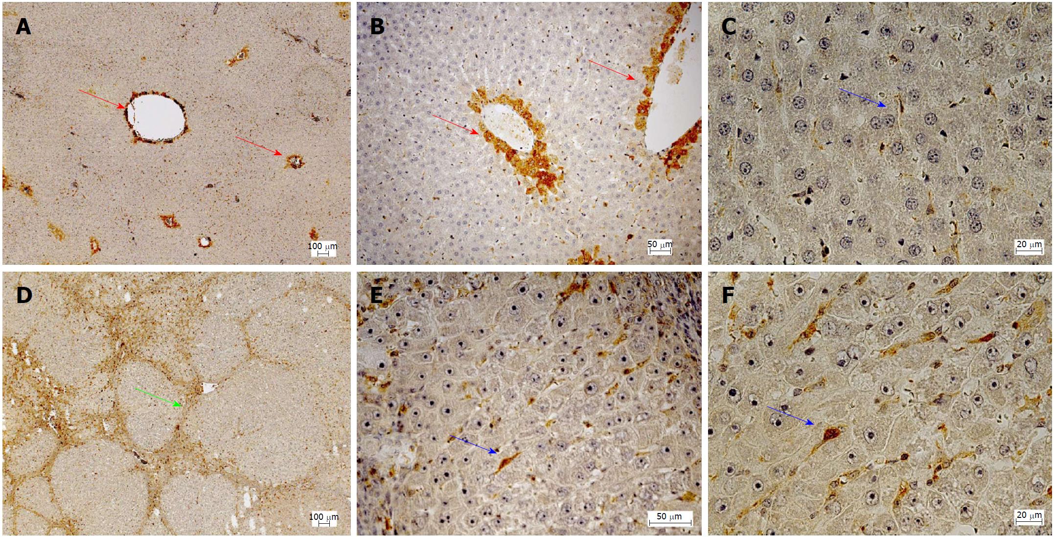
Figure 2 Immunohistochemical localization of phosphodiesterase-5.
A-C: Healthy liver; D-F: Cirrhotic liver. Red arrows: Localization in zone 3 hepatocytes; Blue arrows: Localization in perisinusoidal cells; Green Arrow: Fibrous septa.
- Citation: Schaffner D, Lazaro A, Deibert P, Hasselblatt P, Stoll P, Fauth L, Baumstark MW, Merfort I, Schmitt-Graeff A, Kreisel W. Analysis of the nitric oxide-cyclic guanosine monophosphate pathway in experimental liver cirrhosis suggests phosphodiesterase-5 as potential target to treat portal hypertension. World J Gastroenterol 2018; 24(38): 4356-4368
- URL: https://www.wjgnet.com/1007-9327/full/v24/i38/4356.htm
- DOI: https://dx.doi.org/10.3748/wjg.v24.i38.4356


