Copyright
©The Author(s) 2018.
World J Gastroenterol. Sep 21, 2018; 24(35): 4036-4053
Published online Sep 21, 2018. doi: 10.3748/wjg.v24.i35.4036
Published online Sep 21, 2018. doi: 10.3748/wjg.v24.i35.4036
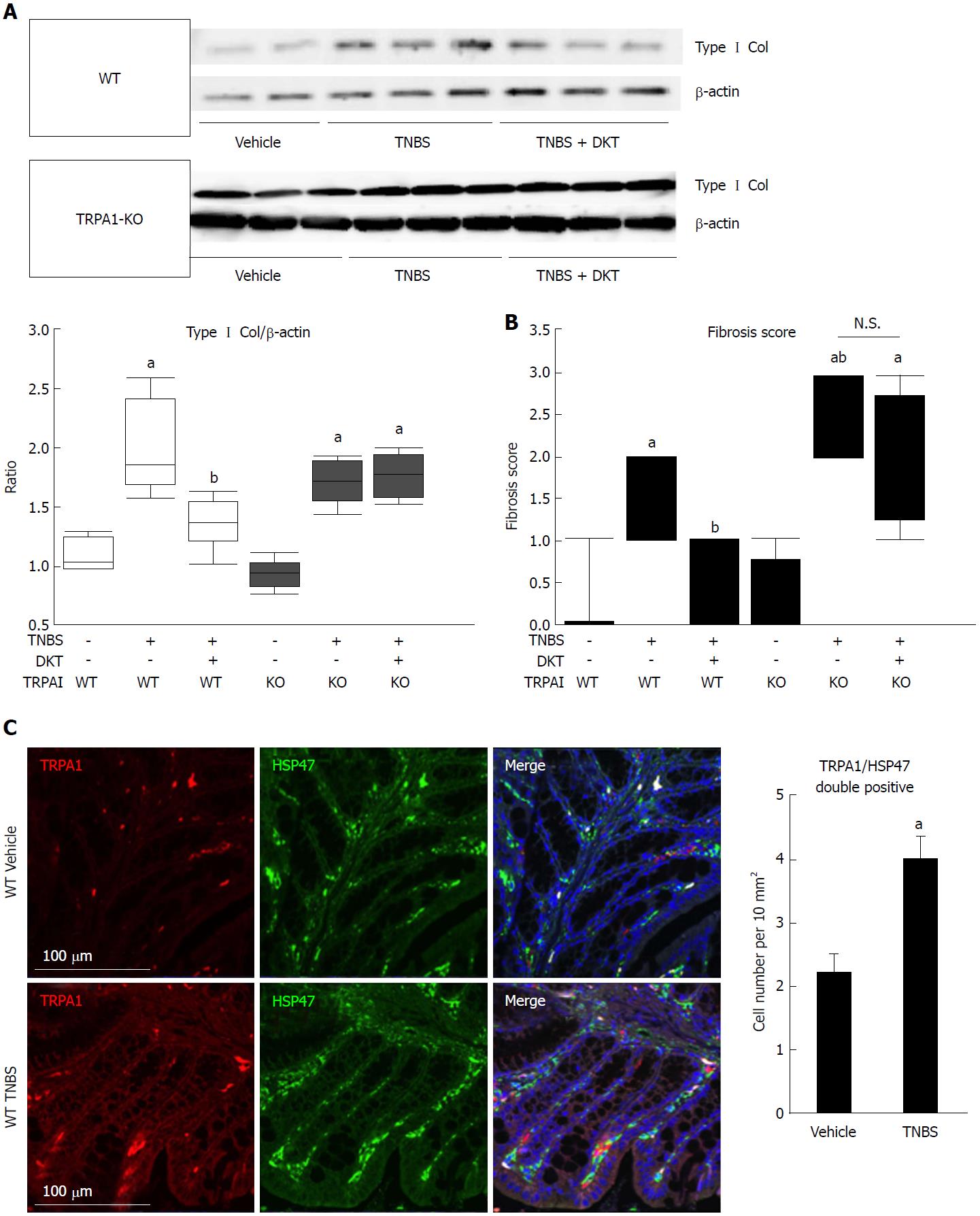
Figure 2 Fibrotic changes in wild-type and transient receptor potential ankyrin 1-knockout CRISPR mice treated with 2, 4, 6-trinitrobenzenesulfonic acid and/or Daikenchuto.
A: Type I collagen expression was examined by western blotting. The ratio of changes in each experimental condition is shown in the middle lower-left graph. aP < 0.01 vs vehicle control, bP < 0.01 vs TNBS (n = 6); B: Fibrosis scores based on histological examination ranging from 0 (no fibrosis) to 3 (severe fibrosis) are shown as the dot-plot graph. The leftmost circles (n = 8) show absolutely zero values. Values are the means ± SD; C: Co-localization of TRPA1 and HSP47 proteins in the mouse intestine. WT vehicle control, WT TNBS: Immunostaining images of mouse intestine stained with anti-TRPA1 (red) antibody, anti-HSP47 (green), and DAPI (blue). Cell number per 10 mm2 is summarized in right graph, aP < 0.05 vs vehicle control (n = 6). TNBS: 2, 4, 6-trinitrobenzenesulfonic acid; WT: Wild-type; HSP47: Heat shock protein 47; TRPA1: Transient receptor potential ankyrin 1; DAPI: 4',6-diamidino-2-phenylindole.
- Citation: Hiraishi K, Kurahara LH, Sumiyoshi M, Hu YP, Koga K, Onitsuka M, Kojima D, Yue L, Takedatsu H, Jian YW, Inoue R. Daikenchuto (Da-Jian-Zhong-Tang) ameliorates intestinal fibrosis by activating myofibroblast transient receptor potential ankyrin 1 channel. World J Gastroenterol 2018; 24(35): 4036-4053
- URL: https://www.wjgnet.com/1007-9327/full/v24/i35/4036.htm
- DOI: https://dx.doi.org/10.3748/wjg.v24.i35.4036


